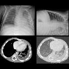
Serothorax


Pleural
effusion • Pleural effusion - Ganzer Fall bei Radiopaedia

Pleural
effusion • Large pleural effusion - infant - Ganzer Fall bei Radiopaedia

Pleural
effusion • Pleural effusion - small - Ganzer Fall bei Radiopaedia

Pleural
effusion • Pleural effusion - infant - Ganzer Fall bei Radiopaedia

Pleural
effusion • Pleural effusion - supine - Ganzer Fall bei Radiopaedia

Pleural
effusion • Right pleural effusion and TIPS - Ganzer Fall bei Radiopaedia

Pleural
effusion • Massive pleural effusion with mediastinal shift - Ganzer Fall bei Radiopaedia

Pleural
effusion • Loculated pleural effusion - Ganzer Fall bei Radiopaedia

Pleural
effusion • Cardiogenic pulmonary edema - Ganzer Fall bei Radiopaedia

Pleural
effusion • Pleural effusion - Ganzer Fall bei Radiopaedia

Pleural
effusion • Small right pleural effusion - Ganzer Fall bei Radiopaedia

Pleural
effusion • Anechoic pleural effusion (ultrasound) - Ganzer Fall bei Radiopaedia

Pleural
effusion • Pleural effusion - Ganzer Fall bei Radiopaedia

Pleural
effusion in an upright position. The fluid is under the lung at the base of the thorax.

Subpulmonic
effusion • Subpulmonic effusion - Ganzer Fall bei Radiopaedia

Pleural
effusion • Pleural metastases from melanoma - Ganzer Fall bei Radiopaedia

Pleural
effusion • Pleural effusion - Ganzer Fall bei Radiopaedia

Pleural
effusion • Meniscus (photo) - Ganzer Fall bei Radiopaedia

Pleural
effusion • Malignant pleural effusion - Ganzer Fall bei Radiopaedia

Round
atelectasis • Pleural thickening: illustrations - Ganzer Fall bei Radiopaedia

Tumor and
tumorlike conditions of the pleura and juxtapleural region: review of imaging findings. Diagnosis: primary effusion lymphoma. Technique: contrast-enhanced chest CT. Description: A 41-year-old man with a history of HIV, HHV8, and EBV presented with dyspnea and chest pain. The initial CT examination (a, b) shows a prominent unilateral pleural effusion posterior to the right lung. A combination of imaging and clinical findings was suggestive of primary effusion lymphoma, which was histopathologically confirmed. Follow-up CT after treatment (c, d) shows a small residual right-sided pleural effusion

Infant with
cough and feverCXR PA and left lateral decubitus shows a left lower lobe infiltrate with a free flowing pleural effusion, confirmed on the axial CT with contrast of the chest which showed no enhancement of the pleura to suggest empyema.The diagnosis was bacterial pneumonia with a reactive pleural effusion.

Mediastinalshift
nach rechts bei großem Pleuraerguss links: Beachte den Verlauf der Trachea.


Pleuraerguss
in der Computertomographie axial Weichteilfenster rechte Pleurahöhle (links im Bild). Die Flüssigkeit sammelt sich beim liegenden Patienten dorsal. Die CT Dichte liegt bei Werten gering über 0 Hounsfield-Einheiten.

Newborn
immediately status post repair of left congenital diaphragmatic hernia. CXR AP (left) shows the hypoplastic lung bud which cannot immediately expand to fill the hemithorax in the apex of the left hemithorax and therefore there is also air in the left pleural space. Note that this is not a pneumothorax and should not be drained via a chest tube. CXR AP obtained 2 days later (right) shows the left pleural space is now filled with fluid rather than air, and again this should not be drained by a chest tube. As the lung bud expands, the pleural effusion will decrease in size.The diagnosis was development of a left pleural effusion to fill the potential space in the left hemithorax after repair of a left congenital diaphragmatic hernia.
 Assoziationen und Differentialdiagnosen zu Pleuraerguss:


