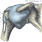
shoulder instability
Glenohumeral instability is the tendency of the glenohumeral joint to sublux or dislocate due to loss of its normal functional or anatomical stabilizers.
Clinical presentation
Glenohumeral instability can be divided into:
- static
- lack of alignment at rest position, which can be depicted using diagnostic imaging studies
- causes include chronic rotator cuff tear and severe osteoarthritis (OA)
- dynamic: lack of alignment during movement or weight-bearing; its etiology can be
- traumatic (TUBS, Traumatic Unilateral dislocations with a Bankart lesion requiring Surgery): most commonly due to episode(s) of anterior joint dislocation and typically associated injuries
- atraumatic (AMBRI, atraumatic, multidirectional, bilateral, rehabilitation, and occasionally requiring an inferior capsular shift): associated with increased capsular laxity, glenohumeral hypermobility and spontaneous dislocation
Glenohumeral instability can also be categorized according to the pattern of instability:
- anterior
- by far the most common, accounting for up to 95% of all cases
- also known as TUBS (see above)
- most frequently due to prior anterior shoulder dislocation
- usually results from anterior glenolabral injury, particularly from disruption of the anterior band of the inferior glenohumeral ligament (IGHL) e.g. Bankart lesion
- posterior
- rare
- also most frequently due to posterior shoulder dislocation
- usually results from posterior glenolabral injury, particularly from disruption of the posterior band of the inferior glenohumeral ligament (IGHL) e.g. reverse Bankart lesion, or disruption of the posterior labrum and/or glenoid rim e.g. Bennett lesion
- associated with glenoid dysplasia and retroversion
- multidirectional
- also known as AMBRI (see above)
- usually not due to previous dislocation, but rather congenital joint capsule laxity
- often bilateral
- superior
- usually associated with multidirectional
As a result of this greater mobility, a number of secondary changes may become evident, including:
- subacromial spur formation
- hypertrophy of the greater tuberosity
- coracoacromial ligament hypertrophy
These changes, in turn, may lead to shoulder impingement.
Risk factors
- previous traumatic dislocation (especially anterior shoulder dislocation)
- athletes (throwing, swimming, tennis)
- congenital
- dysplastic glenoid
- medial anterior shoulder capsular insertion (type III)
- absent or small glenohumeral ligaments
- congenital laxity of capsule or ligaments
Radiographic features
Plain radiograph
Typical bone injuries may be visible, especially in case of anterior instability, but often the radiographs look normal.
CT
CT is superior in visualizing bony injuries of the humeral head and glenoid rim in case of traumatic instability. In atraumatic instability, the findings are often non-specific.
MRI, arthro-MR, arthro-CT
MRI and arthrographic studies are very accurate in showing chondral and labral injuries (such as Bankart lesion, ALPSA, GLAD and HAGL, as well as their counterparts in posterior instability). Visualization of the Perthes lesion may be improved by the use of the ABER position. In multidirectional instability, a circumferential labral tear is often present.
Treatment and prognosis
In general, both anterior and posterior instability requires surgical repair and strengthening of the capsule.
Multi-directional instability is usually treated conservatively with rotator cuff strengthening exercises .
Siehe auch:
- Buford-Komplex
- hintere Schulterluxation
- SLAP-Läsion
- Bankart-Läsion
- Schulterluxation
- ALPSA-Läsion
- vordere Schulterluxation
- humeral avulsion of the glenohumeral ligament
- Schulter Anatomie
- Perthes lesion
- Labrumläsion Schulter
- POLPSA lesion
- Glenohumerale Bänder
- Impingement der Schulter
- glenolabral articular disruption (GLAD)
- posterior glenohumeral instability
- anterior glenohumeral instability
und weiter:

 Assoziationen und Differentialdiagnosen zu glenohumerale Instabilität:


