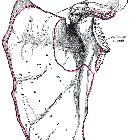
subacromial impingement


Subacromial impingement is by far the most common form of shoulder impingement and occurs secondary to attrition between the coracoacromial arch and the underlying supraspinatus tendon or subacromial bursa, leading to tendinopathy and bursitis respectively.
Pathology
Etiology
- acromial shape
- os acromiale
- type III acromion
- low lying acromion
- downsloping lateral acromion
- acromial spur
- acromioclavicular joint degenerative disease
- coracoacromial ligament ossification or thickening
- shoulder instability
- post-traumatic deformity
- supraspinatus overdevelopment
- chronic bursitis
Radiographic features
Primarily, subacromial impingement is a clinical diagnosis and one should not make a diagnosis or exclude it solely based on imaging. However, imaging has an important role in supporting the diagnosis, finding the possible cause as well as sequelae of impingement.
Static imaging modalities such as MRI and radiographs occasionally depict reduced subacromial distance as indirect evidence:
- type III acromion
- os acromiale
- osteophytes extruding from the acromioclavicular joint inferiorly
- subacromial-subdeltoid bursitis
- lateral acromial slope/tilt
- downsloping lateral acromion
Anecdotal experience also suggests that slight contact between the coracoacromial arch and the subacromial bursa can occur in healthy individuals; yet, significant contact or snapping between these two structures are not common in the absence of symptoms and suggest clinically relevant impingement .
Ultrasound
Dynamic ultrasound may depict abnormal contact between the coracoacromial arch and peritendinous tissue during shoulder abduction; however, dynamic diagnosis at ultrasound is not free of controversy: although earlier studies have demonstrated thickening of the subacromial bursa following shoulder abduction in symptomatic shoulders, a more recent investigation found no significant difference in the degree of bursal gathering in impingement patients compared with healthy volunteers .
Complications
- rotator cuff tear
- subacromial bursitis
- bicipital tendinitis
Related pathology
Less common types of shoulder impingement include:
- subcoracoid impingement: affects subscapularis
- posterosuperior impingement: involves infraspinatus
Siehe auch:
- Klassifikation der Akromiontypen nach Bigliani
- Os acromiale
- posterosuperior impingement
- Bursa subacromialis
- Musculus infraspinatus
- Musculus subscapularis
- Musculus supraspinatus
- Schultersonographie
- Rotatorenmanschette
- spinoglenoidale Zyste
- Impingement der Schulter
- glenohumerale Instabilität
- Akromionsporn
- subcoracoidales Impingement
- type III and type IV acromions
und weiter:


 Assoziationen und Differentialdiagnosen zu subakromiales Impingement: