Soft tissue chondroma of the Hoffa fat pad


Soft tissue chondroma of the Hoffa (infrapatellar) fat pad is a rare form of chondroma. It is considered by some to be the end-stage of Hoffa disease of the infrapatellar fat pad.
Clinical presentation
The condition usually presents in adults with chronic anterior knee pain as well as a hard swelling inferior to the patella.
Pathology
The pathogenesis of this condition is unclear but several theories exist. It may occur as a result of metaplasia from mesenchymal cells due to acute or repetitive trauma related to hyperextension of the knee and subsequent inflammation and hemorrhage .
Microscopic appearance
- lobulated mass of hyaline cartilage
- endochondral ossification in the central areas
- variable areas of:
- fibrocartilage
- myxoid tissue
- fibroadipose tissue
The variety of tissues present accounts for its heterogeneous and variable MRI appearance.
Radiographic features
Plain radiograph
Arc-like calcifications and ossified areas in the infrapatellar fat pad, corresponding to variable endochondral ossification predominant in the central areas of the lobulated mass.
Ultrasound
- lobular mass with acoustic shadowing
CT
- nodular partially calcified cartilaginous mass
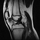
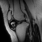
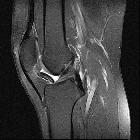
MRI
Features include
-
T1
- mass in the Hoffa fat pad
- generally low signal
- isointense to muscle
- some dark areas signifying calcification
- T2: high signal in the cartilaginous components
- PD: as above with T1, plus high intensity signal areas corresponding to medullary bone
- GRE: prominent signal voids within a nodular mass
Differential diagnosis
Possible considerations include
- synovial chondromatosis
- synovial hemangioma
- soft tissue chondrosarcoma
Practical points
- it is important not to rely solely on MRI or US imaging features alone as the appearances can be variable
- contrast enhancement can occur and does not necessarily imply malignancy
- it is important to correlate the MRI findings with plain radiographs or CT
Siehe auch:


 Assoziationen und Differentialdiagnosen zu Weichteilchondrom des Hoffa'schen Fettkörpers: