spinal pilocytic astrocytoma

Although rare, pilocytic astrocytomas are the most common spinal cord tumors in the pediatric population.
This article specifically relates to spinal pilocytic astrocytomas. For a discussion on intracranial pilocytic astrocytomas refer to pilocytic astrocytoma. For a general discussion on spinal astrocytomas refer to spinal astrocytoma.
Epidemiology
Spinal pilocytic astrocytomas occur most commonly in children and young adults, with 75% of cases occurring in the first two decades of life .
There is no gender predilection.
Associations
There is an association with NF1.
Clinical presentation
Most spinal cord pilocytic astrocytomas are large at presentation as they grow slowly and symptoms evolve over months-years.
Clinical presentation is similar to that of other intramedullary spinal tumors, with pain, weakness and sensory changes common.
Pathology
Pilocytic astrocytomas are benign (WHO grade I) lesions.
Pathology reveals reactive astrocytes with biphasic morphology (loose glial components and compact piloid regions), eosinophilic granular bodies, Rosenthal fibers and hyalinised vessels. There is a lack of mitotic figures and other high-grade features .
Radiographic features
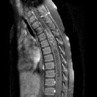
Pilocytic astrocytomas most commonly occur in the thoracic region, followed by the cervical region and less frequently the lumbar region. Some tumors can extend from the brain stem into the cervical cord . Holocord presentation may occur and is more common with pilocytic astrocytomas than other types of astrocytoma.
CT
Hypodense cyst-like component combined with a soft-tissue mural nodule .
MRI
Like other types of astrocytoma, pilocytic astrocytomas typically have an eccentric location within the spinal cord. The most common location is the dorsal portion of the spinal cord .
Fusiform enlargement of the spinal cord is typically seen. Unlike higher grade astrocytomas, pilocytic astrocytomas are almost always well-defined and tend to displace rather than infiltrate the cord .
Associated cysts and syringomyelia are particularly common with pilocytic astrocytomas . Like other types of spinal astrocytoma, hemorrhage is uncommon . Surrounding vasogenic edema is rarely present .
Typical signal characteristics:
- T1: isointense to hypointense
- T2: hyperintense
- T1 C+ (Gd): most enhance but to a lesser degree compared to intracranial pilocytic astrocytomas. The pattern of contrast enhancement is variable
In larger tumors, signal intensity might be heterogeneous due to the intermixture of solid and cystic tumor parts .
Treatment and prognosis
Surgery with the aim of gross total resection is the treatment of choice. The role of radiotherapy is still under debate .
Spinal pilocytic astrocytomas are associated with an excellent prognosis. Ten and 20-year survival rates of between 80 and 100% have been reported in patients who underwent gross total resection. For those with incomplete tumor excision, the 20-year survival rate ranges from 70-80%. Recurrence occurs in 5.4% of totally resected tumors .
Differential diagnosis
- spinal astrocytoma (non-pilocytic)
- difficult to distinguish
- both are seen in patients with NF1
- ependymoma
- patients are usually older
- virtually all enhance strongly
- central location within the spinal cord
- hemorrhage is common
- ganglioglioma
- mixed signal intensity on T1 weighted images
- calcification is common (areas of low signal with blooming on GRE)
- hemangioblastoma
- patients are usually older
- focal flow voids especially in larger lesions
- surrounding edema is usually seen
- hemosiderin capping may be present
- paraganglioma
- patients are usually older
- usually located inferior to the conus
- flow voids are typically seen along the surface of and within the tumor nodule
- hemorrhage is common, leading to a ‘cap sign’
- Intramedullary metastases
- patients are usually older
- prominent edema commonly surrounds the tumor nodule
Siehe auch:
- Pilozytisches Astrozytom
- Ependymom
- Gangliogliom
- Paragangliom
- Hämangioblastom
- spinales Astrozytom
- intramedullary metastases (spinal)
und weiter:


 Assoziationen und Differentialdiagnosen zu spinales Pilozytisches Astrozytom:


