superior labral anterior posterior tear


Superior labral anterior posterior (SLAP) tears are injuries of the glenoid labrum, and can often be confused with a sublabral sulcus on MRI.
Pathology
SLAP tears involve the superior glenoid labrum, where the long head of biceps tendon inserts. They can extend into the tendon, involve the glenohumeral ligaments or extend into other quadrants of the labrum. Unlike Bankart lesions and ALPSA lesions, they are uncommonly (20%) associated with shoulder instability .
Etiology
In the acute setting, they are most frequently seen in falls onto an outstretched arm or in throwing sports athletes.
Classification
The most widely used system for classification of SLAP tears was originally described by Snyder who on the basis of arthroscopic findings, described four patterns of labral injury:
- type I: fraying of the superior labrum free margin
- commonly asymptomatic
- often due to degeneration
- type II: detachment of the superior labrum and biceps anchor from the underlying superior glenoid at the chondrolabral junction (undercuts the anchor)
- in younger patients (<40 years of age) these are associated with Bankart lesions
- in older patients (>40 years of age) they are associated with rotator cuff tears
- seen in overhead athletes
- type III: bucket handle tear of the superior labrum without extension into the long head of biceps tendon
- type IV: bucket handle tear of the superior labrum with extension into the long head of biceps tendon
Beyond these four original types, multiple additional types have been described, although their clinical relevance is controversial. Other described types include :
- type V: anterior-inferior Bankart lesion in continuity with a type II SLAP lesion
- type VI: combination of a type II SLAP lesion and an unstable labral flap either anterior or posterior
- type VII: type II SLAP lesion with extension to the capsule and the middle glenohumeral ligament (MGHL)
- type VIII: type IIB SLAP lesion with posterior labral extension
- type IX: complete or almost complete circumferential detachment of the labrum from the glenoid
- type X: superior labral tear in combination with extension to the rotator cuff interval or the superior glenohumeral ligament or the coracohumeral ligament
Radiographic features
MR arthrogram
The investigation of choice is an MR arthrogram, which is variably reported as having accuracies of 75-90%, although distinguishing between subtypes can be difficult.
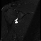
High signal (fluid on T2WI or arthrographic contrast on T1WI) is seen extending into the superior labrum, and tracking into the labrum, and sometimes into the biceps tendon is the characteristic finding.
Treatment and prognosis
- type I tears are usually asymptomatic and do not require treatment
- type II tears require surgical reattachment
- type III tears usually require resection of the bucket handle tear
Differential diagnosis
- sublabral sulcus: SLAP type II tears can appear similar to a normal sublabral sulcus or normal glenoid articular hyaline cartilage which extends beneath the labrum; the following features help distinguish a tear from the latter two
- high T2 signal or contrast curves laterally
- high signal width within labrum >2 mm
- high signal or contrast extends posteriorly to the biceps anchor
- double Oreo cookie sign
- sublabral foramen: SLAP tear type II, in which the labrum is avulsed from the underlying glenoid can look similar to a sublabral foramen (a variant of normal), but can be distinguished from the latter by observing high signal extending between the glenoid and labrum posterior to the attachment of the biceps tendon
- Buford complex
- normal anatomical variant in which there is a congenital absence of the labrum between the 1-3 o'clock positions
Siehe auch:
- sublabrales Foramen
- Labrumläsion Schulter
- Pseudo-SLAP lesion
- Glenoid labrum variants
- intraartikuläre Varianten der langen Bizepssehne
- aufgespaltene lange Bizepssehne
und weiter:


 Assoziationen und Differentialdiagnosen zu SLAP-Läsion:

