talocalcaneal coalition


Talocalcaneal coalition is one of the two most common sub-types of tarsal coalition, the other being calcaneonavicular coalition. It accounts for 45% of all tarsal coalitions, and although all three facets of the talocalcaneal joint can be involved, the middle facet is most commonly involved.
Radiographic features
As with any coalition it may be bony, cartilaginous or fibrous. Talocalcaneal coalition often requires cross-sectional imaging for accurate diagnosis.
Plain radiograph
Plain film findings include:
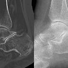
- C-sign
- best assessed on a lateral ankle radiograph
- posterior continuity of the talus and sustentaculum tali
- sensitivity: 50%
- specificity: 90%
- talar beak sign
- best seen on the lateral ankle radiograph
- prominent beak at the anterior aspect of the talus
Secondary radiographic features that suggest the diagnosis include close apposition of the middle facet of the talocalcaneal (subtalar) joint or non-visualization of the middle articular facet . Sclerosis around the articular margins of the talocalcaneal joint may also occur.
CT
At CT, coronal reformats are usually the best to appreciate the coalition. Bony coalition is seen as a complete bar of bone between the talus and calcaneus. In non-osseous coalition, there is usually irregularity of the articular surface, narrowing of the joint space and subchondral sclerosis.
MRI
MRI is more helpful for the assessment of cartilaginous or fibrous coalitions and also demonstrates bone marrow and soft-tissue edema.
Nuclear medicine
On bone scan there is Increased radionucleotide uptake at the site of the coalition may occur due to altered biomechanics at the joint although this is a non-specific finding.
Siehe auch:
- talar beak sign
- Tarsale Koalition
- Kalkaneonavikulare Koalition
- C-Zeichen Talokalkaneare Koalition
- Arthrose unteres Sprunggelenk
- dysplastic sustentaculum tali
- absent middle facet sign
und weiter:


 Assoziationen und Differentialdiagnosen zu Talokalkaneare Koalition: