Talus

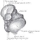

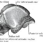
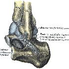


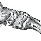
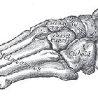
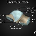

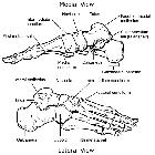

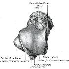
The talus is a tarsal bone in the hindfoot that articulates with the tibia, fibula, calcaneus, and navicular bones. It has no muscular attachments and around 60% of its surface is covered by articular cartilage.
Gross anatomy
The talus has been described as having three main components: head, neck, and body. It is an irregular saddle-shaped bone.
The talar body has a curved smooth trochlear surface also termed the talar dome, which is covered with hyaline cartilage and convex from front to back. The medial and lateral surfaces articulate with the medial malleolus (of the tibia) and lateral malleolus (of the fibula) respectively. The lateral articular surface is large and projects more inferiorly. The lower part of the lateral surface forms a bony projection called the lateral process which supports the lower portion of the lateral articular facet. The posterior aspect has a backward and medially facing posterior process, which has a lateral and medial tubercle separated by a groove for the tendon of flexor hallucis longus.
The talar head is the part that articulates with the navicular bone. On its inferior aspect, this is continuous with three articular facets that are separated by smooth ridges. There are anterior and middle facets which articulate with corresponding facets on the calcaneus. There is another facet, medial to the above facets, for articulation with the spring ligament.
The talar head and body are connected by the talar neck, which is inclined downwards distally and medially.
The inferior surface of the talar neck has a deep groove, the sulcus tali, that passes obliquely forward and expands from medial to lateral. It forms the tarsal sinus with the calcaneal sulcus of the calcaneum. Posterior to the sulcus tali is a large facet that articulates with the posterior talar articular facet of the calcaneus.
Articulations
- superiorly through the talar dome to form the mortise joint of the ankle with the tibia and fibula
- inferoposteriorly: large oblique facet that is concave articulates with the calcaneus to form the talocalcaneal joint
- anteroinferiorly: two facets for articulation with the calcaneus to form part of the talocalcaneonavicular joint
- talar head (domed articular surface) with the navicular bone (circular depression on the posterior surface)
Attachments
Musculotendinous
No muscles originate or insert on the talus.
Ligamentous
- anterior talofibular ligament
- posterior talofibular ligament
- talocalcaneal ligaments
- tarsal sinus ligaments
- cervical ligament
- talocalcaneal interosseous ligament
- deltoid ligament
- dorsal talonavicular ligament
Blood supply
- posterior tibial artery into the medial side of body and sinus
- anterior tibial artery/dorsalis pedis artery into head and neck
- peroneal artery into the lateral side of the body and sinus
The vascular supply to the talus is considered tenuous due to the lack of muscular attachment to the bone .
Innervation
- deep peroneal nerve
- tibial nerve
- saphenous nerve
- sural nerves
Variant anatomy
- talocalcaneal coalition
- os talotibiale - rare accessory bone present on the dorsal aspect of the talar dome, directly anterior to the talocrural joint
- the posterior process of the talus may not be fused to the central portion of the body, resulting in an os trigonum
- type I - complete separation from the talus
- type II - connected to the lateral tubercle through hyaline cartilage
- type III (Stieda process) - lateral tubercle appears elongated as it projects posteriorly
- congenital vertical talus
Related pathology
- osteochondral defect of the talar dome
- avascular necrosis of the talus
- os trigonum syndrome
- fractures
- talar dislocations
Siehe auch:
- Processus lateralis tali Fraktur
- Talusfraktur
- Fußwurzelknochen
- Talusnekrose
- Talus verticalis
- Kugeltalus
- Talusspalte
- Talus obliquus
- Talus secundarius
- Rückfuß
und weiter:

 Assoziationen und Differentialdiagnosen zu Talus:


