Teratom des Thymus

School ager
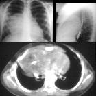
who fell on to her right shoulderCXR AP+lateral shows a large anterior mediastinal mass. Axial CT with contrast of the chest shows a heterogenous mass containing low density fat and high density calcium.The diagnosis was teratoma of the thymus.

School ager
with respiratory distress and an anterior mediastinal mass. Specimen radiograph shows densely calcified rectangular structures in the resected thymic mass which turned out to be teeth.The diagnosis was thymic teratoma.

Role of
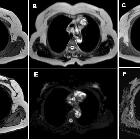
different imaging modalities in the evaluation of normal and diseased thymus. MRI chest in a 45-year-old female patient shows a rather well-defined superior anterior mediastinal lobulated soft tissue lesion eliciting heterogenous low T1 (a) and mixed signal in T2WI signals (b), with areas of restricted diffusion in out-of-phase image (d) compared to in-phase (b, c) image. It elicits diffusion restriction (e, f) with ADC value = 0.8. Histopathological confirmation revealed terato-dermoid tumor
Teratom des Thymus
Siehe auch:

 Assoziationen und Differentialdiagnosen zu Teratom des Thymus:


