thoracic outlet syndrome


Thoracic outlet syndrome (TOS) refers to a group of clinical syndromes caused by congenital or acquired compression of the brachial plexus or subclavian vessels as they pass through the superior thoracic aperture .
Clinical presentation
Clinical presentation will depend on the structure compressed and the degree of compression:
- neurogenic thoracic outlet syndrome
- most common (90-95%)
- brachial plexus compression results in pain, paresthesia and/or numbness of the upper limb
- venous thoracic outlet syndrome
- second most common
- subclavian vein compression causes upper limb swelling and pain and may result in venous thrombosis (Paget-Schroetter syndrome)
- some can present with symptoms of intermittent venous compression in the absence of thrombosis (McCleery syndrome)
- arterial thoracic outlet syndrome
- rarest form (less than 3% of cases)
- subclavian artery compression causes ischemia with coolness, pallor, claudication, paresthesia and decreased upper limb pulses
- usually consists of two components:
- damage to the subclavian artery at the level of the first rib
- distal embolic phenomena
Combined neurovascular symptoms and signs may be present. The findings are exacerbated by certain arm positions and maneuvers, particularly with the arms raised (abducted) above the head.
Pathology
There are three common sites of compression:
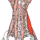
- scalene triangle: between scalenus anterior and scalenus medius muscles
- costoclavicular space: between the clavicle and 1st rib
- subpectoral space: between pectoralis minor and coracoid process
The scalene triangle is defined by the first rib and the anterior and middle scalene muscles and is the most medial compartment. The subclavian artery and branches of the brachial plexus pass through the borders of this triangle while the subclavian vein passes anterior to it.
Etiology
- scalenus anticus syndrome: abnormal insertion of scalenus anterior onto 1st rib (commonest cause)
- congenital cervical rib
- bony abnormality of 1st rib or clavicle (congenital anomaly, malunited fracture, callus, Paget disease, tumor)
- elongated C7 transverse process
- muscle hypertrophy
- fibrous bands
- supraclavicular tumor or lymphadenopathy
Radiographic features
Plain radiograph
Chest radiography is typically performed to exclude an underlying bone abnormality.
Ultrasound / CT / MRI / Angiography
Imaging with ultrasound, contrast-enhanced CT, MRI or conventional angiography is useful for detecting vascular thoracic outlet syndrome (e.g. arterial and/or venous compression). Imaging is performed with the patient’s arms both in the raised (abducted) and neutral (adducted) positions for comparison.
MR imaging is useful in patients with neurogenic thoracic outlet syndrome particularly in evaluating the brachial plexus and surrounding structures.
Imaging findings of thoracic outlet syndrome include :
- neurogenic thoracic outlet syndrome
- bone and soft-tissue abnormalities
- loss of fat about brachial plexus with abduction
- edema in brachial plexus
- venous thoracic outlet syndrome
- bone and soft-tissue abnormalities
- axillosubclavian vein thrombosis
- enlarged collaterals
- fixed axillosubclavian vein stenosis at the site of dynamic narrowing
- axillosubclavian vein narrowing with abduction
- arterial thoracic outlet syndrome
- bone and soft-tissue abnormalities
- axillosubclavian artery aneurysm or pseudoaneurysm
- arterial thrombus
- distal emboli
- enlarged collaterals
- fixed axillosubclavian artery stenosis at site of dynamic narrowing
- axillosubclavian artery narrowing with abduction
Treatment and prognosis
Treatment of arterial thoracic outlet syndrome is surgical intervention. Treatment is required to treat or prevent acute thromboembolic events.
Treatment of venous thoracic outlet syndrome depends primarily on the presence and extent of associated venous thrombosis and may include anticoagulation, thrombolysis, or surgical decompression.
History and etymology
The term "thoracic outlet syndrome" is thought to have been coined by R M Peet et al. in 1956 .
Siehe auch:
- Morbus Paget des Knochens
- Halsrippe
- Gabelrippe
- Musculus scalenus anterior
- Paget-von-Schroetter-Syndrom
- vaskuläre Kompressionssyndrome
- Venous thoracic outlet syndrome
- Srb-Anomalie
- Musculus scalenus medius
und weiter:


 Assoziationen und Differentialdiagnosen zu Thoracic-outlet-Syndrom: