Thrombose der tiefen Hirnvenen


Deep cerebral vein thrombosis is a subset of cerebral venous thrombosis involving the internal cerebral veins, often coexisting with cortical vein thrombosis or dural venous sinus thrombosis, and with different clinical presentations relying on which segment is involved.
As such please refer to the cerebral venous thrombosis article for a general discussion.
Epidemiology
Deep cerebral vein thrombosis may occur at any age. A female predilection exists, and pregnant/peripartum women as well as women on the contraceptive pill are over represented. Elderly patients are more likely to be affected. Please refer to the generic article for a broad discussion on epidemiology and risk factors: cerebral venous thrombosis.
Clinical presentation
Presentation is highly variable, but compared to run-of-the-mill dural venous sinus thrombosis is usually shorter and more rapidly progressive to profound deficit . Presentation can include: nausea, vomiting, headache, focal neurological deficit, seizure, hemiparesis, aphasia, and coma.
Pathology
Please refer to the generic article for a broad discussion on pathology and risk factors: cerebral venous thrombosis.
Radiographic features
Typically the thalami are bilaterally edematous with potential superimposed venous infarction and hemorrhage, although occasionally the findings will be markedly asymmetric or unilateral . For unknown reasons the right side is more often involved than the left when the involvement is asymmetric or unilateral.
CT
Non-enhanced CT is usually the first imaging investigation performed given the non-specific clinical presentation in this cases. When not associated with venous hemorrhage or infarction, it can be hard to make the diagnosis. Potential findings include:
- dense clot sign
- cortical edema
- peripheral hemorrhage (cortical linear density or gyriform heterogeneous hemorrhage)
With contrast administration, especially with a CT venogram, then a filling defect in the veins and sinuses is sought.
MRI
MRI is able to both visualize the clot as well as the sequelae.
- T1: The acute clot is isointense becoming hyperintense in the subacute phase; see aging blood on MRI
- T2
- acute clot is hypointense on T2 (this can mimic a flow void)
- hyperintense swelling of thalami and basal ganglia
- MR venography: will demonstrate lack of flow
Treatment and prognosis
For general discussion on treatment please refer to the parent article: cerebral venous thrombosis.
Compared to dural venous sinus thrombosis, deep cerebral venous thrombosis, especially when the internal cerebral veins are involved, carries a poorer prognosis .
Practical points
- midline and bilateral infarction areas (e.g. involving thalami or parasagittal cortex)
- cortical or peripheral hemorrhage, especially when heterogeneous and gyriform
- cortical edema
- direct signs of a thrombus (e.g. dense clot sign, cord sign)
Siehe auch:
- Sinusthrombose
- Altersbestimmung Blutung MRT
- zerebrale Venenthrombose
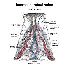
- internal cerebral veins
- venous infarction
und weiter:


 Assoziationen und Differentialdiagnosen zu Thrombose der tiefen Hirnvenen: