Trachealtumoren


A case of
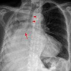
tracheal obstruction presenting as COPD. Initial PA Chest X-Ray on presentation, demonstrating a tracheal stenosis at the level of the clavicle and a hyper inflated chest with flattened hemi-diaphragms, consistent with COPD.

A case of
tracheal obstruction presenting as COPD. Coronal thoracic CT demonstrating a marked narrowing of the trachea, below the level of the thyroid, with a 3.5 cm inferior extension.

A case of
tracheal obstruction presenting as COPD. Transverse thoracic CT demonstrating a significant reduction in tracheal luminal diameter with the absence of any focal lung lesions, suggestive of a primary tumour. The oesophagus appears compressed. Mediastinal lymph nodes are not seen.

About a
submucosal tracheal tumor. Computed tomography showed a circular mass in the lower trachea (arrow), with extra tracheal development.

About a
submucosal tracheal tumor. Spiral computed tomography showed that the tumor extended just across the carina (arrow).

A rare case
report of Extraskeletal Ewing"s sarcoma of the trachea. Plain CT Thorax shows a well defined polypoidal soft tissue density lesion in the left anterolateral wall of the trachea at T2 vertebral level with significant luminal narrowing and mediastinal extension

A rare case
report of Extraskeletal Ewing"s sarcoma of the trachea. CT Thorax lung window shows no abnormalities in lung parenchyma.
Tumoren der Trachea
Trachealtumoren
Siehe auch:
- Lungenkarzinom
- Trachea
- Trachealstenose
- Raumforderungen der Trachea
- Tumoren des Tracheobronchialsystems
- Plattenepithelkarzinom der Trachea
- tracheobronchiale Metastasen
- adenoid-zystisches Karzinom des Tracheobronchialbaums
- gestielte intratracheale Läsionen
- Hämangiom der Trachea
- tracheal leiomyosarcoma
- Granulom der Trachea
- Polypen der Trachea
und weiter:

 Assoziationen und Differentialdiagnosen zu Tumoren der Trachea:


