tracheobronchial injury

Anesthetic
management of tracheal laceration from traumatic dislocation of the first rib: a case report and literature of the review. Preoperative evaluation of Tracheobronchial lacerations by a high-resolution CT. a Sagittal CT image of the chest showing the posterior tracheal wall laceration up to 59.81 mm below the glottis and 63.76 mm above the carina. b, c Axial CT image of the chest showing the bone shadow in the trachea; the residual largest cavity of the trachea on the left was 5.33 mm in diameter and 6.66 mm on the right

Clinical
features and management of closed injury of the cervical trachea due to blunt trauma. CT scan of the neck, showing disruption of the cervical trachea and widespread subcutaneous emphysema.

School ager
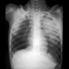
in a motor vehicle accident with respiratory distressCXR AP shows air in the superior mediastinum and neck, airspace disease in the left upper lobe, and a small amount of air in the left pleural space.The diagnosis was tracheobronchial injury of the left mainstem bronchus with pneumomediastinum and a small left pneumothorax.
Tracheobronchial injury is a serious but uncommon manifestation of chest trauma. It is usually a fatal injury with only a small percentage of patients making it to hospital. Given the magnitude of force required to injure the major airways, there are often multiple chest injuries and other body regions affected.
Pathology
The cervical trachea is the most injured site in penetrating injury , whereas injury to the distal trachea (within 2.5cm of the carina) is seen most commonly following blunt chest trauma . The site of injury can be identified in 70% of patients with CT .
Radiographic features
Plain radiograph
- subcutaneous emphysema in the chest and/or neck
- pneumothorax
- pneumomediastinum
- endotracheal tube balloon
- overinflation
- abnormal shape
- herniation into the airway defect
CT
Features include :
- disruption of the tracheal or bronchial cartilage rings
- irregularity of the airway wall
- focal thickening of the tracheal or bronchial wall
- laryngeal disruption
- massive pneumomediastinum (sometimes despite appropriate pleural space decompression with intercostal catheters)
- fallen lung sign (first described in 1970 ): rare
Complications
If left untreated, late complications can occur such as :
- recurrent infection (pneumonia)
- bronchial or tracheal stenosis
- bronchiectasis
Differential diagnosis
- tracheal diverticulum: usually on the right and at the level of the superior thoracic aperture
Siehe auch:
und weiter:

 Assoziationen und Differentialdiagnosen zu tracheobronchiale Verletzungen:


