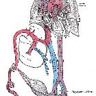
umbilical artery


The umbilical artery gives rise to both a nonfunctional remnant of the fetal circulation and an active vessel giving supply to the bladder. In the adult, the obliterated area of the vessel is identifiable as the medial umbilical ligament and the patent segment is the superior vesical artery.
Summary
- origin: anterior division of the internal iliac artery
- location: abdominal wall and pelvis
- supply: placenta in the fetus, superior bladder, ureter, ductus deferens
- main branches: obliterated umbilical artery, superior vesical artery
Gross anatomy
Origin
The umbilical artery originates from the anterior division of the internal iliac artery. In the fetus, it travels within the umbilical cord to the placenta and hence is the communication to the maternal circulation.
After the umbilical cord has been cut at birth, clots form within the vessel and it obliterates. Its remnant can be found in the posterior aspect of the anterior abdominal wall as the medial umbilical ligament, which courses superomedially to the umbilicus, within a fold of peritoneum called the medial umbilical fold. It is a paired structure and lies lateral to the median umbilical ligament (the urachal remnant) and medial to the lateral umbilical fold, which contains the inferior epigastric vessels.
Branches
Within the fetus, the artery terminates in the placenta as multiple chorionic vessels.
In the adult, there are two vessels which the umbilical artery gives rise to; these are not branches but are actually a direct continuation of the umbilical artery:
- obliterated umbilical artery: this is the distal part of the umbilical artery that thromboses soon after birth and hence becomes obliterated; it may be found as a remnant seen in the internal aspect of the anterior abdominal wall
- superior vesical artery: the persistent proximal portion of the umbilical artery; runs along the side wall of the pelvis to its supply at the superior bladder as well as adjacent ureter and ductus deferens
Supply
In the fetus, the paired umbilical arteries travel within the umbilical cord to carry deoxygenated blood from the fetus to the mother.
In the adult, the superior vesical artery gives vascular supply to the superior bladder, the portion of the ureter next to this and the ductus deferens.
Siehe auch:
- Dopplersonographie der Nabelarterie
- singuläre Nabelschnurarterie (sNSA)
- Ligamentum teres vesicae
- reversal of umbilical artery end diastolic flow
- absent umbilical artery end diastolic flow
und weiter:

 Assoziationen und Differentialdiagnosen zu Nabelarterie:


