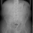
Urethrakonkrement im Penis

Urethral
calculi with a urethral fistula: a case report and review of the literature. a Computed tomography scan of the soft tissue in the penis showing multiple calculi in the distal urethra. b Computed tomography scan of the pelvis showing a thickened urinary bladder and Foley’ s catheter through the abdominal wall. c Computed tomography scan of the scrotum showing a purulent infection and its spread in the perineal fascia space

Urethral
calculi presenting with acute urinary retention. Radiodense opacity at the penis level.

Urethral
calculus • Penile urethral calculus - Ganzer Fall bei Radiopaedia

Urethral
calculus • Penile urethral calculus - Ganzer Fall bei Radiopaedia

Urethral
calculi presenting with acute urinary retention. Axial reconstruction at the penis base level shows two impacted urethral calculi (yellow arrow).

Urethral
calculi presenting with acute urinary retention. Sagittal reconstruction shows two impacted urethral calculi (yellow arrow) and another one of smaller dimension in the bladder lumen (white arrow). Note the Foley catheter adequately placed (green star).

Urethral
calculus • Urethral stone - Ganzer Fall bei Radiopaedia
Urethrakonkrement im Penis
Urethrakonkrement Radiopaedia • CC-by-nc-sa 3.0 • de
Urethral calculi are an uncommon type of urolithiasis, accounting for ~1% of all urinary tract stones.
Epidemiology
They almost all occur in males with two peak incidences - one in childhood and the other at 40 years .
Clinical presentation
Most commonly acute lower urinary tract symptoms and/or urinary retention.
Pathology
Urethral calculi are most commonly calcium oxalate (~85%) and can be either :
- primary: arising de novo secondary to other pathologies such as diverticuli, strictures, neurogenic bladder or foreign bodies
- secondary: originate in the upper urinary tract (much more common)
Location
Most impact in the prostatic urethra although ~40% (range 30-50%) are found in the anterior urethra .
Radiographic features
Almost all (98-100%) of urethral stones are reported to be radiopaque but most are small and only faintly radiopaque and up to 60% will be missed .
Differential diagnosis
- pelvic phlebolith
- prostatic calcification: transrectal ultrasound can be helpful for differentiation
- Coaptite injection
Siehe auch:

 Assoziationen und Differentialdiagnosen zu Urethrakonkrement im Penis: