vanishing white matter disease
Vanishing white matter disease (VWM), also known as childhood ataxia with central hypomyelination (CACH), is an exceedingly rare entity only fully described in 1997, but due to its name sometimes over-represented in differentials for white matter disease.
Epidemiology
Most cases are encountered in young patients with onset in early childhood or adolescence being typical . A number of adult patients have however been described .
Presentation is preceded by a minor head trauma or infection resulting in episodic deterioration followed by incomplete and slow recovery. With repeated episodes patients accumulate neurological impairment eventually succumbing to the disease .
Clinical presentation
Currently the diagnosis is made on the basis of clinical and MRI appearances, as no established diagnostic laboratory tests exist .
Diagnostic criteria have been proposed :
- cerebellar ataxia
- spasticity
- optic atrophy (not always)
- epilepsy (not always)
- motor functions disproportionately affected
For the diagnosis to be made, all four criteria should be identified .
There is some evidence that vanishing white matter disease is an autosomal recessive condition, and it is believed that the culprit gene resides on chromosome 3q27 .
Pathology
Histology reveals :
- rarefaction of the white matter (more severe in the deeper white matter)
- microcystic change in the periventricular white matter
- spongiform change in the arcuate fibers and the corpus callosum
- neuronal loss is not present
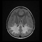
Radiographic features
MRI
MRI is the modality of choice for assessing cases of possible vanishing white matter disease, and other white matter disease.
White matter is diffusely involved, extending from periventricular white matter to the subcortical arcuate fibers. Over time the white matter vanishes replaced by near-CSF intensity fluid (i.e. it attenuates on FLAIR) .
Cerebellar atrophy is variably present and ranges form severe to mild and typically involves the vermis .
MRS is useful. In affected white matter, only lactate and glucose peaks remain. In unaffected grey matter, the trace is near-normal with small glucose and lactate peaks .
Treatment and prognosis
Although the disease is spontaneously remitting, each episode results in accumulation of further injury. No treatment has been identified and death usually occurs within months to years (range: few months to 14 years) .
Differential diagnosis
When a potential case is encountered, it is unusual for the reading radiologist to have had any significant personal experience with the disease, and as such a differential needs to be entertained, including :
- leukodystrophies, especially: Alexander's disease
- deterioration is not episodic
- patients are usually macrocephalic
- white matter is affected in a fronto-occipital gradient
- histology: Rosenthal fibers are present
Siehe auch:
und weiter:


 Assoziationen und Differentialdiagnosen zu leukoencephalopathy with vanishing white matter: