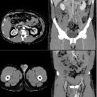
Varikozelenembolisation

Zustand nach
Embolisation der Vena testicularis links bei Varikozele. Die Röntgenübersichtsaufnahme des Abdomens zeigt die längs aneinander gereihten, gewundenen Coils in Projektion auf den Psoas.

Zustand nach
Embolisation der Vena testicularis links bei Varikozele. In der Computertomographie axial sieht man im Verlauf der Vene vor dem Psoas die metalldichten Coils mit entsprechenden Artefakten.

Varicocele
embolization • Varicocele embolization - Ganzer Fall bei Radiopaedia

Varicocele
embolization • Varicocele embolization with polidokanol and coil - Ganzer Fall bei Radiopaedia

Varicocele
embolization • Amyand hernia with acute appendicitis - Ganzer Fall bei Radiopaedia

Role of
varicocele sclerotherapy in the management of benign prostatic hyperplasia and its associated lower urinary tract symptoms (pilot study). Diagram showing A normal, B pathological and C post sclerotherapy testicular-prostatic venous changes with emphasis on the hydrostatic pressure changes for blood column lengths (EIV = external iliac vein, IIV = internal iliac vein, CIV = common iliac vein)

Role of
varicocele sclerotherapy in the management of benign prostatic hyperplasia and its associated lower urinary tract symptoms (pilot study). A prostatic capsular contrast ‘blush’ (long arrows) following spermatic venous opacification confirms the backdoor phenomenon [7] “Copyright (2022) Wiley. Used with permission from (Gat and Goren, Benign prostatic hyperplasia: Long-term follow-up of prostatic volume reduction after sclerotherapy of the internal spermatic veins, Andrologia, Wiley)” (pp = pampiniform plexus)
 Assoziationen und Differentialdiagnosen zu Varikozelenembolisation:


