gonadale Venen


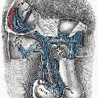

The gonadal veins are paired structures that drain the gonads in males and females. In males it is called the testicular vein (or internal spermatic vein) and in females it is called the ovarian vein. The gonadal veins ascend with the gonadal arteries in the abdomen along the psoas muscle anterior to the ureters. Like the suprarenal veins each side drains differently:
- the left gonadal vein drains into the left renal vein
- the right gonadal vein drains directly into the inferior vena cava
Gross anatomy
Ovarian veins
Ovarian veins emerge from the ovary as a plexus (pampiniform plexus) in the mesovarium and suspensory ligament. Two veins emerge from the plexus and ascend with the ovarian artery, usually merging into a single vein before termination.
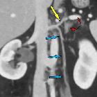
The right ovarian vein arises from the right pampiniform plexus (which is continuous with the uterine plexus) and lies lateral to the right ureter. It ascends anterior to psoas and parallels the right ureter. It crosses the ureter anteromedially halfway between bifurcation of the IVC and the point in which it joins the anterolateral inferior vena cava (IVC).
The left ovarian vein ascends similarly into the abdomen but drains into the left renal vein .
Testicular veins
The venous circulation of the male gonads comprises two intrascrotal networks; a deep and superficial venous network. The deep network drains the testis, epididymis, and ductus deferens. The superficial network drains the veins of the scrotum.
Veins of the testes and epididymis form the pampiniform plexus. which ascends to form four veins at the level of the superficial inguinal ring and a single testicular vein at the level of the deep inguinal ring. It ascends through the inguinal canal in the spermatic cord.
The testicular vein ascends in the retroperitoneum on psoas major. Along its course there are variable communications with retroperitoneal veins, abdominal wall veins and renal capsular veins. The left testicular vein invariably drains into the left renal vein. The right testicular vein usually drains into the IVC just below the renal vein, but sometimes drains into the right renal vein.
Variant anatomy
- duplication is common, more often on the left (~13%) than on the right (~2%)
- both gonadal veins draining into the left renal vein
- in doubled IVC the left gonadal vein drains into the left IVC
- partially divided gonadal veins with the gonadal arteries running through
- gonadal veins may receive a duodenal or suprarenal vein
- gonadal veins draining into common iliac veins
- right gonadal vein drains into the right renal vein
- may be in the form of a plexus
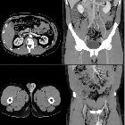
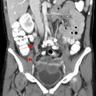
Radiographic features
CT
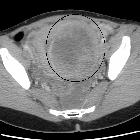
- the ovarian veins are frequently used to identify the ovaries; they are useful landmarks particularly for determining the origin of a pelvic mass
- the ovarian vascular pedicle sign is present in 92% of ovarian masses; direct joining of an asymmetrically enlarged gonadal veins with a pelvic mass indicates that the ovary is the organ of origin
- the ovarian vein is best visualized at level of the origin of the inferior mesenteric artery where it is surrounded by retroperitoneal fat and in the pelvis medial to external iliac vessels
- testicular vein normally measures 1-3 mm in diameter
Related pathology
- ovarian vein phlebolith
- ovarian vein reflux and pelvic congestion syndrome
- ovarian vein thrombosis
- ovarian vein stenosis and occlusion
- ovarian vein syndrome
- ovarian venous varix
- varicocele
Siehe auch:
- Ovarialvenenthrombose
- Phlebolithen in Ovarialvene
- Varicocele der Vena ovarica
- Vena ovarica
- ovarian vein syndrome
- Vena testicularis
- Embolisation Vena ovarica
- Varikozelenembolisation
- Vena uterina
- Ovarialvenenthrombose rechts
- ovarian vein congestion
und weiter:


 Assoziationen und Differentialdiagnosen zu gonadale Venen: