Vesical fungus ball

Fungusball in der Harnblase
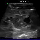
Vesical fungus ball
Fungusball im Harntrakt Radiopaedia • CC-by-nc-sa 3.0 • de
Fungal balls of the urinary tract, also known as fungal bezoars or mycetomas of the urinary tract, are a rare manifestation of funguria, usually candiduria.
Epidemiology
While candiduria may be seen in approximately 20% of hospitalized patients , development of fungal balls is considered very uncommon, although the exact incidence is unknown. They are more commonly seen in the neonatal and elderly demographics .
Risk factors
Fungal balls are generally seen in the presence of at least one of the following :
- diabetes mellitus
- immunocompromised state (e.g. post-transplant patients, HIV with poor virological suppression)
- indwelling catheterization
- anatomical urinary tract anomalies
- urinary retention (e.g. bladder obstruction, neurogenic bladder)
- prolonged antibiotic therapy
- concurrent malignancy
Clinical presentation
Fungal balls of the urinary tract are usually diagnosed in the context of a symptomatic urinary tract infection or urosepsis, and are not symptomatic themselves unless they cause obstruction . If obstructive, they may contribute to renal colic-type pain, development of or worsening of pyelonephritis, and a post-renal acute kidney injury .
Pathology
Etiology
The etiology of fungal balls is not clearly understood, however they are thought to be an agglutination of :
- mycelia
- mucoid debris
- fragments from papillary necrosis
- lithiasic debris
- gas
The most commonly implicated pathogen is Candida albicans, however many other fungal isolates have been described in the literature, including other Candida spp., Aspergillus spp., Rhizopus oryzae, and Geotrichum candidum .
Location
Fungal balls can exist anywhere along the urinary tract, however are most commonly described as existing within the renal pelvis .
Radiographic features
Fungal balls are often seen in the setting of cystitis, pyelonephritis, papillary necrosis, and/or evidence of urinary tract obstruction (e.g. hydronephrosis, hydroureter) .
Plain radiograph
Plain radiography is often unremarkable, however unusual locules of gas or calcification may be seen within the urinary system possibly indicative of underlying fungal ball .
Fluoroscopy
Cystography and pyelography may reveal contrast filling defects where the fungal balls lie .
Ultrasound
Generally, fungal balls are appreciated as mobile, rounded, heterogeneously hypoechoic masses, although hyperechoic masses have also less frequently been described . No evidence of vascularity is seen within the mass on a Doppler study .
CT
Fungal balls have a heterogeneous soft-tissue density on CT without contrast-enhancement, but may also have regions of gas or calcification . They are rounded and not attached to the walls of the urinary tract, with a thin rim of urine often appreciable around the mass . CT urography (e.g. CT intravenous pyelogram) reveals filling defects, similar to fluoroscopy .
MRI
Morphological findings are identical to those seen on CT . Signal characteristics of fungal balls have been rarely described, but include :
- T1: isointense to renal parenchyma
- T2: hyperintense to renal parenchyma
Similar to other modalities, MR urography reveals filling defects .
Treatment and prognosis
Treatment is typically with local and/or systemic antifungal therapy, such as fluconazole or amphotericin B . If refractory to pharmacotherapy, fungal balls may be surgically removed .
Differential diagnosis
Depending on the modality, the following may be considered:
- hematoma
- urolithiasis
- tumor
- inflammatory debris
Siehe auch:


 Assoziationen und Differentialdiagnosen zu Fungusball in der Harnblase: