zystische Metastasen

Branchial
cleft anomalies: a pictorial review of embryological development and spectrum of imaging findings. Forty-year-old man presenting with a persistent level II neck lump. Axial T2-weighted imaging demonstrates a complex cystic lesion posterior to the left angle of the mandible (white arrow). Asymmetrical soft tissue was also appreciated in the left fossa of Rosenmüller (asterisk). A second branchial cleft cyst was considered in the differential, but the diagnosis was finally confirmed to be nasopharyngeal carcinoma with cystic metastases

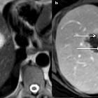
Cystic liver
lesions: a pictorial review. Cystic metastasis of a mucinous tumour of the rectum in a 72-year-old male. The hyperintensity on axial T2-weighted magnetic resonance imaging a is marked but the multiple tiny septa (arrows) incompletely enhanced on portal venous phase (b) are sufficient to reach a conclusive diagnosis in this the context

Cystic liver
lesions: a pictorial review. Pure cystic metastasis of a neuroendocrine tumour in a 69-year-old female. The hyperintensity on axial T2-weighted magnetic resonance imaging (a) is marked and there is no enhancement on arterial (arrow) or portal venous (arrow-head) phase (b, c)

Cystic liver
lesions: a pictorial review. Metastatic pancreatic neuroendocrine tumour in a 60-year-old male. a Axial computed tomography on portal venous phase shows a lobulated hypodense cystic nodule in segment V with enhanced mural nodule and septa (arrow), very hyperintense on T2-weighted magnetic resonance imaging (b). A second cystic subcapsular nodule is visible in segment VI

Cystic liver
lesions: a pictorial review. Cystic metastasis from malignant melanoma in a 72-year-old female. a, b Axial computed tomography on arterial and portal venous phases shows unique hypoattenuating (20 Hounsfield Units) nodule of the right liver lobe, too small to be characterised. c Contrast-enhanced ultrasonography displays an enhanced wall as a “rim” (arrows)

A practical
approach to cystic lung disease on HRCT. Multiple cystic lesions are present in the left lung (arrow) with a small pneumothorax (arrowhead). The patient had a known history a previous oropharyngeal squamous cell carcinoma and the lesions were confirmed as cystic squamous cell carcinoma metastases

Thyroglossal
duct pathology and mimics. Necrotic metastatic lymphadenopathy (primary later found to be tongue base squamous cell carcinoma). Axial contrast-enhanced CT image illustrates an irregularly thick-walled cystic mass anterior to the left thyroid cartilage (arrow). Careful search in other areas of the neck demonstrate another similar appearing cystic lymph node adjacent to the right thyroid cartilage (arrowhead)

Zystische
Metastase eines papillären Schilddrüsenkarzinoms: Der große zystische Befund rechts zervikal zeigte mehrere solide, deutlich kontrastmittelaufnehmende Anteile. Neben dem Hauptbefund waren weitere Lymphknoten auffällig. MRT T2 axial und koronar, T1 axial und T1 axial FatSat Kontrastmittel. Sonografie des Lymphknotens und des Schilddrüsenknotens.
 Assoziationen und Differentialdiagnosen zu zystische Metastasen:

