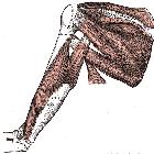
rotator cuff tears


Rotator cuff tears are one of the most common causes of shoulder pain mostly in older patients.
Clinical presentation
Prevalence of tear increases with age. Most significant findings are impingement and "arc of pain" sign (pain while lowering the abducted arm) . Supraspinatus weakness, night pain and weakness of external rotation (seen in infraspinatus tear) may also be present.
Pathology
Etiology
Important causes include:
- traumatic
- acute
- chronic repetitive
- degenerative
- subacromial impingement
- tendon degeneration
- hypovascularity
Location
Rotator cuff tears can affect each one of the rotator cuff muscles or more than one. The supraspinatus muscle is most commonly affected, followed by infraspinatus, subscapularis and teres minor muscles .
- tendon insertion: often degenerative
- critical zone: degenerative or trauma related
- myotendinous junction: often trauma-related, infraspinatus muscle most often affected
Subtypes
A modification of the original Codman classification (1930) may be used to categorize tears:
- full-thickness rotator cuff tear
- partial-thickness rotator cuff tear
- intrasubstance tear: not in communication with the joint surface or with the bursal surface of the tendon
- articular-sided tear
- rim rent tear: articular surface tear of the footprint
- with tendon delamination or interstitial tear; if the gap is filled with fluid then it is called cleavage tear of the rotator cuff
- bursal-sided tear
- critical zone tear: partial or full-thickness
Radiographic features
Exact features depend on the type of tear. General features include
Plain radiograph
Typically, these are normal in acute tears with chronic tears showing degenerative-type changes :
- may show a decreased acromiohumeral interval
- <7 mm on true AP shoulder radiograph in chronic tears
- <2 mm on an 'active abduction' view in acute tears
- may show decreased supraspinatus opacity and decreased bulk due to fatty atrophy in chronic tears
- humeral subluxation superiorly may be seen in chronic tears
- may show features of subacromial impingement
- spur formation on the undersurface of acromioclavicular joint
- acromion with an inferolateral tilt seen on outlet view (i.e. modified 'Y' view)
- type III acromion
- secondary degenerative changes: sclerosis, subchondral cysts, osteolysis, and notching/pitting of greater tuberosity
Ultrasound
Ultrasound may have up to 90% sensitivity and specificity. It can also reveal other mimics like tendinosis, calcific tendinitis, subacromial-subdeltoid bursitis, greater tuberosity fracture, and adhesive capsulitis.
Full-thickness tears extend from bursal to the articular surface, while partial-thickness tears are focal defects in the tendon that involve either the bursal or articular surface. Full-thickness appear on ultrasound as hypoechoic/anechoic defects in the tendon. Due to the fluid replacing tendon, cartilage shadow gets accentuated giving a double cortex or cartilage interface sign. Also, due to the defect, overlying peribursal fat dips down into the tendon gap, creating a sagging peribursal fat sign .
Direct signs are:
- non-visualization of the supraspinatus tendon
- hypoechoic discontinuity in the tendon
Indirect signs are:
- double cortex sign
- sagging peribursal fat sign
- compressibility
- muscle atrophy
Secondary associated signs are:
- cortical irregularity of greater tuberosity
- shoulder joint effusion
- fluid along the biceps tendon
- fluid in the axillary pouch and posterior recess
MRI
Full-thickness tears are easier to diagnose on MRI than partial-thickness tears . Hyperintense signal area within the tendon on T2W, fat-suppressed, intermediate-weighted and GRE sequences, usually matches to fluid signal. They extend from articular to the bursal surface. The presence of a tendon defect filled with fluid is the most direct sign of rotator cuff tear. Tendon retraction may also be present, which can be graded using the Patte classification. Indirect signs on MRI are - subdeltoid bursal effusion, medial dislocation of biceps, fluid along biceps tendon, and diffuse loss of peribursal fat planes. Muscle atrophy and fatty replacement is seen in chronic cases and can be graded using the Goutallier classification, or assessed with the tangent sign or scapular ratio .
Partial-thickness tears extend to either the bursal or articular surface, and sometimes they are interstitial that means confined within the tendon. Retraction of tendinous fibers from the distal insertion into the greater tuberosity may also be considered partial tear.
Chronic tears show signs of extensive use or repetitive stress, which can be reflected by intramuscular cystic changes and tendinosis in the remaining tendon. There are often risk factors for subacromial impingement present for example degenerative changes at the acromioclavicular joint or acromioclavicular joint cysts.
MR arthrography may enhance the detection of rotator cuff tears, especially full-thickness tears.
Differential diagnosis
General imaging differential considerations include:
- tendinosis/tendinopathy
- calcific tendinitis
- subacromial-subdeltoid bursitis
- greater tuberosity fracture
- adhesive capsulitis
See also
- rotator cuff
- rotator cuff interval
- rotator cuff arthropathy
- Goutallier classification of rotator cuff muscle atrophy
- tangent sign
Siehe auch:
- Humeruskopfhochstand
- Tendinitis calcarea
- Tendinosis calcarea der Rotatorenmanschette
- Patte-Klassifikation der Sehnenretraktion bei Rotatorenmanschettenruptur
- Rotatorenmanschette
- rotator cuff interval
- frozen shoulder
- Ruptur Supraspinatussehne
- partial articular supraspinatus tendon avulsion (pasta) lesion
- rotator cuff repair
- partial rotator cuff tears
- Klassifikation von Partialrupturen der Rotatorenmanschette nach Ausdehnung nach Ellman
- Sonographie Rotatorenmanschettenruptur
- Ruptur der Subscapularissehne
- rotator cuff tear grading
- Retraktionsgrad nach Patte bei kompletten Rotatorenmanschettenrupturen
- Grading Rotatorenmanschettenruptur
- Snyder classification of partial rotator cuff tears
- Rotatorenmanschette Pitfalls
und weiter:

 Assoziationen und Differentialdiagnosen zu Rotatorenmanschettenruptur:


