cytomegalovirus encephalitis


Cytomegalovirus (CMV) encephalitis is a CNS infection that almost always develops in the context of profound immunosuppression.
This article focus on adult infection. CMV is also one of the most frequent prenatal infections, which is discussed separately: congenital CMV infection.
Epidemiology
It is extremely rare to involve immunocompetent individuals.
Clinical presentation
Presentation is variable, however generalized cognitive decline is most frequent. Focal neurological signs and cranial nerve palsies are uncommon to rare.
At autopsy active CMV infection is also seen systemically although clinical manifestations are variable:
- CMV adrenalitis (92%)
- CMV pneumonitis (42%)
- CMV retinitis (58%)
Pathology
Typically, before CMV infections become clinically apparent the patient's CD4 count typically reaches below 50/mm . CMV infects the entire neuraxis (brain, spinal cord, meninges, nerve roots, eye) and therefore has variable presentation.
Etiology
Cytomegalovirus is a DNA virus belonging to the herpesviruses group, and is also known as Human Herpesvirus 5 (HHV-5). It is found in all geographic locations and socioeconomic groups, and infects between 50-80% of adults in the United States as indicated by the presence of antibodies in much of the general population.
Radiographic features
Two most common imaging findings are meningoencephalitis and ventriculitis/ependymitis.
MRI
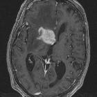
In CMV encephalitis, there is usually only non specific increased T2/FLAIR signal in the white matter. If ventriculitis is also present then enhancement of the ependymal surface and hydrocephalus may also be seen.
high T2 white matter change most prominent in a periventricular distribution
- no enhancement (unless ventriculitis present, in which case 30% or so will enhance)
- no mass effect (often seen with concurrent atrophy)
Appearances are similar to HIV encephalitis, although usually CMV occurs in patients with a longer duration of disease, and lower CD4+ counts.
Differential diagnosis
Other white matter changes in HIV, especially :
- HIV encephalitis
- progressive multifocal leukoencephalitis (PML)
- primary CNS lymphoma: periventricular but has mass effect and enhances
Practical points
Particularly in a profoundly immunocompromised patient, ventriculitis characterized by high T2/FLAIR signal and post contrast enhancement is highly suggestive of CMV infection.
Siehe auch:
- primäres ZNS-Lymphom
- Progressive multifokale Leukenzephalopathie
- Virusenzephalitis
- HIV-Enzephalopathie
- Enzephalitis
- white matter changes in HIV
- Zytomegalievirus
und weiter:

 Assoziationen und Differentialdiagnosen zu Zytomegalievirus Enzephalitis:


