Danis-Weber classification

Weber
classification of ankle fractures • Weber fracture classification (illustration) - Ganzer Fall bei Radiopaedia

Left side
shows removed parts, right side of same JPG is the corresponding X-ray. The eye-catching very long screw was removed 6 weeks after installation, the remaining screws were removed 18 months after installation.

Weber
classification of ankle fractures • Ankle fracture - Weber B - Ganzer Fall bei Radiopaedia

Weber
classification of ankle fractures • Distal fibula fracture - Ganzer Fall bei Radiopaedia

Weber
classification of ankle fractures • Ankle fracture - Weber A - Ganzer Fall bei Radiopaedia

Weber
classification of ankle fractures • Ankle fracture - Weber C - Ganzer Fall bei Radiopaedia

Weber
classification of ankle fractures • Ankle fracture - Weber C - Ganzer Fall bei Radiopaedia

Weber
classification of ankle fractures • Ankle fracture - Weber C - Ganzer Fall bei Radiopaedia

Weber
classification of ankle fractures • Ankle fracture - Weber C - Ganzer Fall bei Radiopaedia
Weber
classification of ankle fractures • Ankle (distal fibular) fracture - Weber C - Ganzer Fall bei Radiopaedia

Weber
classification of ankle fractures • Ankle fracture - Weber B - Ganzer Fall bei Radiopaedia

Weber
classification of ankle fractures • Ankle fracture - Weber B - Ganzer Fall bei Radiopaedia

Weber
classification of ankle fractures • Ankle fracture - Weber B - Ganzer Fall bei Radiopaedia

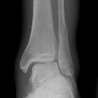
Weber-A-Fraktur:
Frakturlinie an der Spitze der distalen Fibula, deutlich unterhalb der tibiofibularen Syndesmose, hier ohne größere Dislokation.

Weber
classification of ankle fractures • Ankle fracture - Weber A - Ganzer Fall bei Radiopaedia

Weber
classification of ankle fractures • Ankle fracture - Weber A - Ganzer Fall bei Radiopaedia

Weber
classification of ankle fractures • Ankle fracture - Weber A - Ganzer Fall bei Radiopaedia

Weber
classification of ankle fractures • Ankle fracture - Weber A - Ganzer Fall bei Radiopaedia

Weber
classification of ankle fractures • Ankle fracture - Weber A - Ganzer Fall bei Radiopaedia

Weber
classification of ankle fractures • Ankle fracture - Weber A - Ganzer Fall bei Radiopaedia

Weber
classification of ankle fractures • Weber classification of ankle fractures - Ganzer Fall bei Radiopaedia


Weber
classification of ankle fractures • Ankle fracture - Weber C - Ganzer Fall bei Radiopaedia
The Weber ankle fracture classification (or Danis-Weber classification) is a simple system for classification of lateral malleolar fractures, relating to the level of the fracture in relation to the ankle joint, specifically the distal tibiofibular syndesmosis. It has a role in determining treatment.
Classification
- type A
- below the level of the syndesmosis (infrasyndesmotic)
- usually transverse
- tibiofibular syndesmosis intact
- deltoid ligament intact
- medial malleolus occasionally fractured
- usually stable if medial malleolus intact
- type B
- distal extent at the level of the syndesmosis (trans-syndesmotic); may extend some distance proximally
- usually spiral
- tibiofibular syndesmosis usually intact, but widening of the distal tibiofibular joint (especially on stressed views) indicates syndesmotic injury
- medial malleolus may be fractured
- deltoid ligament may be torn, indicated by widening of the space between the medial malleolus and talar dome
- variable stability, dependent on the status of medial structures (malleolus/deltoid ligament) and syndesmosis; may require ORIF
- Weber B fractures could be further subclassified as
- B1: isolated
- B2: associated with a medial lesion (malleolus or ligament)
- B3: associated with a medial lesion and fracture of posterolateral tibia
- type C
- above the level of the syndesmosis (suprasyndesmotic)
- tibiofibular syndesmosis disruption with widening of the distal tibiofibular articulation
- medial malleolus fracture or deltoid ligament injury often present
- fracture may arise as proximally as the level of fibular neck and not visualized on ankle films, requiring knee or full-length tibia-fibula radiographs (Maisonneuve fracture)
- unstable: usually requires ORIF
- Weber C fractures can be further subclassified as
- C1: diaphyseal fracture of the fibula, simple
- C2: diaphyseal fracture of the fibula, complex
- C3: proximal fracture of the fibula
- a fracture above the syndesmosis results from external rotation or abduction forces that also disrupt the joint
- usually associated with an injury to the medial side
History and etymology
This classification was first described by the Belgian general surgeon, Robert Danis (1880-1962), in 1949. It was later modified and popularized by the Swiss orthopedic surgeon, Bernhard Georg Weber (1929-2002), in 1972 .
See also
Siehe auch:
- Weber-B-Fraktur
- Weber-C-Fraktur
- Weber-A-Fraktur
- Pilon tibiale Fraktur
- Ligamentum collaterale mediale
- Ottawa ankle rules
- distale Fibulafrakturen
- trimalleoläre Sprunggelenksfraktur
- bimalleoläre Sprunggelenksfraktur
- Lauge-Hansen classification
- kindliche Sprunggelenksfrakturen
und weiter:


 Assoziationen und Differentialdiagnosen zu Danis-Weber classification:


trimalleoläre
Sprunggelenksfraktur