Osteoradionekrose der Mandibula

Mandibular
osteoradionecrosis • Osteoradionecrosis of mandible with infection - Ganzer Fall bei Radiopaedia

Mandibular
osteoradionecrosis • Mandibular osteoradionecrosis with fracture - Ganzer Fall bei Radiopaedia

Mandibular
osteoradionecrosis • Osteoradionecrosis of the mandible - Ganzer Fall bei Radiopaedia

Mandibular
osteoradionecrosis • Mandibular osteoradionecrosis with pathological fracture - Ganzer Fall bei Radiopaedia

Mandibular
osteoradionecrosis • Mandibular osteoradionecrosis - Ganzer Fall bei Radiopaedia

Mandibular
osteoradionecrosis • Mandibular osteoradionecrosis - Ganzer Fall bei Radiopaedia

Osteoradionecrosis
• Mandibular osteoradionecrosis - Ganzer Fall bei Radiopaedia
Osteoradionecrosis
• Osteoradionecrosis of the mandible - Ganzer Fall bei Radiopaedia

Osteoradionecrosis
• Osteoradionecrosis of the mandible - Ganzer Fall bei Radiopaedia

Radiolucent
lesions of the mandible: a pattern-based approach to diagnosis. CT aspect of osteoradionecrosis 5 years after radiotherapy of a floor of the mouth SCC. a OPT. b Axial bone window CT image. c Three-dimensional reconstruction, posterior view obtained after removing the bony structures of the cervical spine. Characteristic osteolytic bone defect (arrows) with poorly defined partly sclerotic margins and soft tissue ulceration (dashed arrow). Follow-up confirmed absence of tumour

Radiolucent
lesions of the mandible: a pattern-based approach to diagnosis. MRI aspect of osteoradionecrosis 2 years after chemoradiotherapy of a tonsillar SCC. a Axial T1-weighted image. b Contrast-enhanced sagittal oblique T1-weighted image and ADC map (small image in b). Bilateral hypointensity of the bony marrow. Large osseous defect with cortical destruction on the left (arrow in a). Marked, non-specific contrast enhancement, bony destruction and necrotic hypointense sequestrae (arrow in b). Note high ADC value (red circle) measured at 1.53 × 10⁻³ mm2/s suggesting inflammation and absent recurrence. c Macroscopic resection specimen. The necrotic area is indicated by arrows. d Histology (haematoxylin-eosin stain, original magnification 40×): necrotic bone trabeculae with inflammatory infiltrative changes in the bony marrow
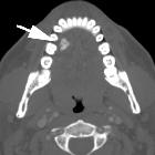
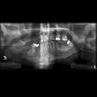
Mandibular osteoradionecrosis (ORN) is more common after radiation therapy for head and neck malignancies due to the superficial position of the mandible, which exposes it to high radiation. The maxilla can also be involved, but this is less frequent.
Epidemiology
Mandibular ORN may occur in ~20% (5-37%) of patients .
Pathology
Mandibular ORN typically occurs in a patient who has received a dose of >60 Gy . Osteoradionecrosis changes may occur within a year of therapy.
Radiographic features
Features include :
- cortical destruction that is ill-defined resulting in a mixed sclerotic-lucent pattern
- sequestration, especially of the buccal bone
- an absence of soft tissue mass is an important feature to differentiate it from neoplastic recurrence but the presence of soft tissue does not exclude ORN
Treatment and prognosis
Conservative treatment is initially medication only (e.g. pentoxifylline, vitamin E) but more severe cases may require hyperbaric oxygen therapy and/or debridement. Some patients will require resection and reconstruction of the mandible .
Complications
- pathological fractures
- infection
- radiation-induced neoplasia
Differential diagnosis
Siehe auch:


 Assoziationen und Differentialdiagnosen zu Osteoradionekrose der Mandibula: