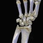
perilunäre Luxation


Perilunate dislocations and perilunate fracture-dislocations are potentially devastating closed wrist injuries that are often missed on initial imaging.
These injuries involve dislocation of the carpus relative to the lunate which remains in normal alignment with the distal radius. They should not be confused with lunate dislocations where the lunate is dislocated in a volar direction and no longer has normal radiolunate articulation.
Epidemiology
Overall, carpal dislocations account for less than 10% of all wrist injuries.
Perilunate dislocations typically occur in young adults with high energy trauma resulting in the loading of a hyperextended, ulnarly deviated hand. Around 60% of perilunate dislocations are associated with a scaphoid fracture which is then termed a trans-scaphoid perilunate dislocation.
Mechanism
Typical history is of a fall onto a dorsiflexed wrist. There may or may not be obvious clinical deformity. Occasionally median nerve injury, arterial compromise or compartment syndrome may be evident due to the dislocation.
Perilunate dislocation involves traumatic rupture of the radioscaphocapitate, scapholunate interosseous and lunotriquetral interosseous ligaments. Mayfield et. al. have proposed a four-stage process to describe perilunar wrist instability (see carpal dislocation) where perilunate dislocation represents stage 2 .
Radiographic features
Imaging plays an essential role in identifying perilunate and other carpal dislocations. Unfortunately, dislocations can often be missed by radiologists. Carpal alignment needs to be carefully assessed on all radiographs. The majority of cases involve dorsal dislocation of the capitate and carpus relative to the lunate which remains in near-normal alignment with the radius. Volar perilunate dislocation is rare (see case 9).
In a trans-scaphoid perilunate dislocation the proximal scaphoid maintains its lunate relationship, and the distal scaphoid and remainder of the carpal bones displace dorsally .
Plain radiograph
- AP radiograph
- dislocation is often overlooked
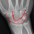
- disruption of the normally smooth line made by tracing the proximal articular surfaces of the hamate and capitate
- increased overlap of lunate and capitate
- piece of pie sign: although also seen in lunate dislocation it may prove very helpful in initial identification of lunate related pathology
- lateral radiograph
- dislocation more easily appreciable
- capitate not sitting within the distal articular 'cup' of the lunate
- line drawn through radius and lunate fails to intersect capitate
- lunate remains in articulation with distal radius (as opposed to lunate dislocation where it is usually in a volar position)
- abnormal scapholunate angle (normal 30-60 degrees, reduced in dorsal perilunate dislocation)
- abnormal capitolunate angle (normal 0-30 degrees, increased in dorsal perilunate dislocation)
Report checklist
In addition to stating that a perilunate dislocation is present, a number of features should be sought and commented upon:
- dislocation
- ensure also that the triquetrum or lunotriquetral ligaments are intact, as if either is disrupted then it is a midcarpal dislocation (stage III carpal dislocation)
- ensure that radiolunate alignment is maintained and that you are not looking at a lunate dislocation (stage IV carpal dislocation)
- associated fractures
- scaphoid (trans-scaphoid-perilunate dislocation)
- present in 60% of cases
- radial styloid
- trapezium
- capitate (transcapitate perilunate dislocation)
- triquetrum (transtriquetral perilunate dislocation)
- scaphoid (trans-scaphoid-perilunate dislocation)
CT
Plain film is normally sufficient to diagnose the dislocation (assuming there is no interpretation error), however, CT plays an important role in assessing for associated occult fractures; the most common and important being scaphoid fracture.
Treatment and prognosis
Untreated there is a high risk of median nerve palsy, pressure necrosis, compartment syndrome, and long-term wrist dysfunction. As with other dislocations, perilunate dislocation should be reduced as soon as possible. Prompt open reduction with the ligamentous repair is necessary.
Despite treatment, the long-term risk of degenerative arthritis is high (~60%). Dissociative and non-dissociative carpal instability can occur with DISI or VISI pattern. Also, there is a higher rate of nonunion of scaphoid fractures when associated with perilunate dislocation than with isolated scaphoid fractures.
Differential diagnosis
The most important differential diagnosis is that of a lunate dislocation which can mimic a perilunate dislocation, especially on AP projection. The key to not confusing the two is the lateral projection:
- in a lunate dislocation, the radiolunate articulation is disrupted and the lunate is dislocated in a palmar direction
- in a perilunate dislocation, the radiolunate articulation is maintained
See also
Siehe auch:
- DISI-Fehlstellung
- PISI-Fehlstellung
- Gilula-Linien
- Scapholunäre Dissoziation
- Lunatumluxation
- Luxationen der oberen Extremität
- perilunäre Luxationsfraktur
- karpale Luxationen
- transscaphoidale perilunäre Luxation
- perilunäre Instabilität
- Mayfield-Klassifikation
- karpale Luxationen und Luxationsfrakturen
- perilunate injuries
und weiter:

 Assoziationen und Differentialdiagnosen zu perilunäre Luxation:


