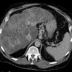
portal-hypertensive Gastropathie

Portal
hypertensive Gastropathie bei Stenose der Pfortader im Rahmen einer chronischen Pankreatitis. Computertomografie in der portalvenösen Phase links axial, rechts koronar: Es zeigt sich die deutliche ödematöse Verdickung der Magenwand mit Hyperämie der Mucosa.

Portal
hypertensive gastroenterocolopathy • Portal hypertensive gastropathy, enteropathy, and colopathy - Ganzer Fall bei Radiopaedia

Portal
hypertensive gastroenterocolopathy • Splenorenal shunt venous aneurysm - Ganzer Fall bei Radiopaedia

Image of
en:portal hypertensive gastropathy seen on endoscopy of the en:stomach. The normally smooth mucosa of the stomach has developed a en:mosaic like appearance, that resembles en:snake-skin. -- Samir 03:52, 14 August 2007 (UTC) en:Category:Endoscopic images
In portal hypertension, chronic portal venous congestion leads to dilatation and ectasia of the submucosal vessels in the stomach (portal hypertensive gastropathy), small bowel (portal hypertensive enteropathy) and/or large bowel (portal hypertensive colopathy). This may result in upper or lower gastrointestinal (GI) bleeding, even in the absence of varices. The bleeding may be acute or chronic but is most commonly chronic low-grade GI blood loss associated with an iron-deficiency anemia.
Radiographic features
Fluoroscopy
Barium studies may show thickening of the mucosal folds and nodular filling defects.
CT
On CT there may be bowel wall thickening and hyperemia which can mimic enterocolitis.
Siehe auch:
und weiter:

 Assoziationen und Differentialdiagnosen zu portal-hypertensive Gastropathie:


