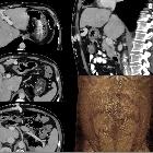
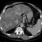
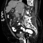
portal-systemic collateral pathways

Caput medusae
bei Pfortaderhochdruck bei Leberzirrhose in der Computertomographie. (sagittal)

Caput medusae
mit Durchtritt oberhalb des Nabels über Verbindungen der rekanalisierten Nabelvene zu den epigastrischen Gefäßen.

Sehr
voluminöse portokavale Umgehungskreisläufe kleinkurvaturseitig am Magen mit Ausdehnung nach intragastral bis knapp submukös.

Fundusvarizen
bei Leberzirrhose mit portaler Hypertension. Computertomographie axial.

Portal
hypertension • Gastroesophageal varices - Ganzer Fall bei Radiopaedia

Portosystemic
collateral pathways • Superior mesenteric venous (SMV) varices - Ganzer Fall bei Radiopaedia

Portal
hypertension • Enlarged paraumbilical vein - Ganzer Fall bei Radiopaedia

Ösophagusvarizen
bei Leberzirrhose mit portaler Hypertension. Computertomographie axial.

Ösophagusvarizen
bei Leberzirrhose mit portaler Hypertension. Computertomographie axial.

Portosystemische
Umgehungskreisläufe über Nabelvenen: Caput medusae bei Leberzirrhose. Computertomographie axial

Portosystemische
Umgehungskreisläufe über Nabelvenen: Caput medusae bei Leberzirrhose. Computertomographie sagittal

Caput medusae
bei Pfortaderhochdruck bei Leberzirrhose in der Computertomographie. (axial)

Caput medusae
bei Pfortaderhochdruck bei Leberzirrhose in der Computertomographie. (axial)

Großkalibrige
portosystemische Umgehungskreisläufe über Venen der Bauchwand. Computertomographie: Rekonstruktion mit Volume-rendering-Technik

Großkalibrige
portosystemische Umgehungskreisläufe über Venen der Bauchwand. Computertomographie: Rekonstruktion mit Volume-rendering-Technik

Großkalibrige
portosystemische Umgehungskreisläufe über Venen der Bauchwand. Computertomographie: Rekonstruktion mit Volume-rendering-Technik

Portosystemic
collateral pathways • Hepatic cirrhosis with portal hypertension and massive ascites - Ganzer Fall bei Radiopaedia
Portosystemic collateral pathways (also called varices) develop spontaneously via dilatation of pre-existing anastomoses between the portal and systemic venous systems. This facilitates shunting of blood away from the liver into the systemic venous system in portal hypertension, as a means for reducing portal venous pressure. However, these are not sufficient for normalizing portal venous pressure.
The main sites of portosystemic collateral pathways are:
- left gastric (see gastric varices)
- left gastric (coronary) vein and short gastric veins to distal esophageal veins
- located between medial wall of gastric body and posterior margin of left hepatic lobe in lesser omentum
- usually accompanied by esophageal/paraesophageal varices (see below)
- esophageal: dilated submucosal venous plexus of distal esophagus (see esophageal varices)
- paraesophageal
- coronary vein to azygos and hemiazygos veins and vertebral venous plexus
- located posterior to esophagus in posterior mediastinum
- perisplenic: splenic vein to left renal vein, traversing splenocolic ligament; may eventuate in spontaneous splenorenal shunt
- retrogastric
- left gastric (coronary) vein or gastroepiploic vein to esophageal or paraesophageal veins
- located in posterior/posteromedial aspect of gastric fundus
- retroperitoneal-paravertebral: colic or mesenteric branches of superior mesenteric vein to retroperitoneal/lumbar veins to the inferior vena cava
- paraumbilical: left portal vein to paraumbilical veins in anterior ridge of falciform ligament
- anterior abdominal wall: paraumbilical and omental veins to subcutaneous periumbilical veins and superior and inferior epigastric veins; collaterals appear to radiate from the umbilicus (caput medusae)
- mesenteric: dilated branches of superior mesenteric vein
- anal canal: superior rectal vein (from the inferior mesenteric vein) to upper anal canal veins (hemorrhoids)
Differential diagnosis
Dilatation of splenic veins at the splenic hilum mimicking splenic hilar varices without splenomegaly may occur in situations such as increased perfusion of splenic tissue associated with an immune response .
Siehe auch:
- rekanalisierte Umbilikalvene
- portale Hypertension
- Caput medusae
- Ösophagusvarizen
- transjugulärer intrahepatischer portosystemischer Shunt
- Abernethy malformation
- Fundusvarizen
- aneurysmal intrahepatic portal-hepatic venous shunt
- intrahepatische Shunts
- angeborener intrahepatischer portosystemischer Shunt
- intrahepatischer portosystemischer Shunt
- splenorenaler Shunt
und weiter:

 Assoziationen und Differentialdiagnosen zu portosystemische Umgehungskreisläufe:
transjugulärer
intrahepatischer portosystemischer Shunt


