Empyema necessitans


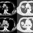
Empyema necessitans (also sometimes spelled as empyema necessitasis) refers to extension of an empyema out of the pleural space and into the neighboring chest wall and surrounding soft tissues.
Pathology
It may either occur due the virulence of the organism or may be facilitated by previous thoracic surgery (e.g. thoracotomy) or trauma allowing infection to track through. It occurs commonly to subcutaneous tissues of the chest wall, but can also spread to involve other sites such as the esophageal, breast, retroperitoneal, peritoneal, pericardial and paravertebral regions. The resultant subcutaneous abscess may eventually rupture through the skin.
Causative organisms
- Mycobacterium tuberculosis: thought to be most common cause and may account for ~70% of cases
- Actinomyces spp.: considered second most common cause (see: thoracic actinomycocis infection)
- Blastomycosis spp.
- Aspergillus spp.
- Nocardia: see pulmonary nocardiosis
- Mucormycosis spp.
- Fusobacterium spp.
Radiographic features
Plain radiograph
Findings on chest radiographs are often non-specific and at times can even be normal. May suggest a soft tissue density in the chest wall.
CT
Chest CT is best at assessing extent of infection out of the thoracic cavity:
- will classically show an empyema (often relatively well-demarcated collection) with extension through the chest wall into another compartment
- adjacent rib destruction may be present
Treatment and prognosis
Management options include closed or open drainage of the pleural space to prevent fibrosis and to facilitate expansion of the lung. Appropriate antibiotic therapy is also a mainstay of treatment .
Differential diagnosis
General imaging differential considerations include:
- malignant pleural-based mass e.g. mesothelioma but will have different clinical context and will have more solid components
- transdiaphragmatic spread of intra- or infra-abdominal infection and/or collection
Siehe auch:
und weiter:

 Assoziationen und Differentialdiagnosen zu Empyema necessitans:


