Kleinhirntumoren
Pilocytic
astrocytoma - MRI appearance. MRI T1W axial image shows a well-defined hypointense lesion with inner hypo-isointense area in left cerebellum.

Pilocytic
astrocytoma - MRI appearance. MRI T2W axial image shows a well-defined hyperintense lesion with inner hypointense area noted in left cerebellum. The lesion shows mild perilesional oedema and causes compression over 4th ventricle resulting in mild hydrocephalus.

Cerebellar
pilocytic astrocytoma: MR spectroscopy. Shows cystic lesion with eccentric enhancing mural nodule in posterior fossa

Cerebellar
pilocytic astrocytoma: MR spectroscopy. T1WI shows hypointense lesion in the posterior fossa

Cerebellar
pilocytic astrocytoma: MR spectroscopy. T2WI shows hyperintense lesion in posterior fossa with effaced 4th ventricle and triventricular hydrocephalus with eccenteric intermediate signal intensity solid component.

Cerebellar
pilocytic astrocytoma: MR spectroscopy. Post contrast T1WI shows peripheral enhancing cystic lesion with eccentric mural nodule.

Cerebellar
pilocytic astrocytoma: MR spectroscopy. MRS shows laclate doublet at 1.3ppm and elevated CHO peak at 3.2ppm and reduced NAA peak and increase cho/Cr and cho/NAA ratio.

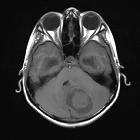
Verkalktes
Meningeom rechts infratentoriell (nicht histologisch bestätigt). Links im Fenster für das Hirnparenchym, rechts Knochenfenster.

Tumor-to-tumor
metastases in Cowden’s disease: an autopsy case report and review of the literature. MRI findings in cerebellar dysplastic gangliocytoma. T1 (a) and T2 (b) weighted magnetic resonance images of the head. A striped pattern is seen in the right hemisphere of the cerebellum (arrowheads). The cerebral aqueduct is compressed

Embryonal
tumor with multilayered rosettes. Infratentorial midline tumour affecting the cerebellum and filling the fourth ventricle. The lesion is solid and hyperattenuating, with punctate calcifications and discrete cystic foci.

Embryonal
tumor with multilayered rosettes. Infratentorial tumour of the midline affecting the cerebellum and the fourth ventricle, with compression and displacement of the brainstem, and a poor cleavage plane with the vermis.

Embryonal
tumor with multilayered rosettes. Presence of low signal elements due to the presence of calcium and blood.

Embryonal
tumor with multilayered rosettes. Heterogeneous enhancement of the tumour is observed.
Kleinhirntumoren
Siehe auch:
- Hirnabszess
- Ependymom
- Kleinhirninfarkt
- Hämangioblastom
- zystische Raumforderung hintere Schädelgrube
- Lhermitte-Duclos-Syndrom
- zerebelläres Gliom
- Cerebellitis
- primäre Hirntumoren
- Pilozytisches Astrozytom des Kleinhirns
und weiter:
- Pilozytisches Astrozytom
- Hirntumoren
- infratentorielles Meningeom
- Meningeom am Tentorium
- Embryonaler Tumor mit mehrreihigen Rosetten
- anaplastisches infratentorielles Ependymom
- Ependymom des Vierten Ventrikels
- Tumoren des vierten Ventrikels
- cerebellar metastases
- infratentoriell supratentoriell
- zystische zerebelläre Raumforderungen
- infratentorielles pilozytisches Astrozytom
- juveniles zerebelläres pilozytisches Astrozytom
- cerebellar medulloblastoma in childhood
- hemispheric medulloblastoma
- zerebelläres Lymphom


 Assoziationen und Differentialdiagnosen zu Kleinhirntumoren:


