Rhabdomyosarkom der Blase

Toddler with
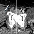
recently noted urinary retention. AP image from the excretory phase of a vintage intravenous pyelogram (above) shows bilaterally normal appearing renal collecting systems and ureters. There is a multilobulated circumferential filling defect in the base of the badder. Axial CT with contrast of the abdomen shows the mass to almost complete fill the base of the bladder.The diagnosis was rhabdomyosarcoma of the bladder.

Preschooler
with a palpable midline abdominal mass. AXR (above left) shows a soft tissue mass in the mid abdomen displacing the bowel loops superiorly. Transverse and sagittal US of the mass (below left) show a solid, homogenous, lobulated mass. Axial CT with contrast of the abdomen (below right) shows a large heterogeneous mass in the mid to left abdomen with a low density center and swirling enhancement. There was a suggestion of direct tumor invasion into the right rectus muscle anteriorly (above right) and the bladder inferiorly.The diagnosis was rhabdomyosarcoma arising from the dome of the bladder.
 Assoziationen und Differentialdiagnosen zu Rhabdomyosarkom der Blase:


