sacral tumours


Epidemiological
characteristics of 1385 primary sacral tumors in one institution in China. Location distribution of chordoma and giant cell tumor. a Chordomas at the Region S1 or S2 or S1-2 accounting for 1.6% (5/316); b Chordomas at the Region S3 or below S3 accounting for 45.9% (145/316); c Chordomas associated with both high and low levels accounting for 52.5% (166/316); d GCT at the Region S1 or S2 or S1-2 accounting for 39.0% (80/205); e GCT at the Region S3 or below S3 accounting for 5.4% (11/205); f GCT associated with both high and low levels accounting for 55.6% (114/205)

Transarterial
embolization (TAE) of sacral giant cell Tumor (GCT) using spherical parmanent embolic material superabsorbant polymer microsphere (SAP-MS). A 68-year-old female with GCT in the sacrum (Case 2). A. On CT, a large osteolytic mass occupied the sacrum (white arrows). B. On CT of same level as Figure 1A about 10 years after initial TAE, the size of the mass decreased and obvious reossification is shown (white arrows).

Große
sakrale Metastase eines Prostatakarzinoms mit großer, ventraler, extraossärer Komponente und Zeichen einer intraläsionalen Einblutung. Oben links Computertomografie, rechts MRT T2-SPIR, unten links T1, rechts T1 FatSat nach Kontrastmittel.

Atypical
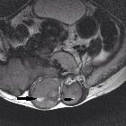
giant sacral myxopapillary ependymoma with massive osteolysis. T2-weighted image in the axial plane: hyperintensity of a mass originating from the sacrum and extending into the presacral space.

Atypical
giant sacral myxopapillary ependymoma with massive osteolysis. Axial T1-weighted image: hypointense mass originating form the sacrum and extendimg into the presacral space.

Atypical
giant sacral myxopapillary ependymoma with massive osteolysis. Hyperintensity on DWI (a) and hypointensity on ADC (b) of a sacral mass, indicating hypercellularity.

Atypical
giant sacral myxopapillary ependymoma with massive osteolysis. Weak contrast enhancement of a sacral mass after Gadolinium administration.

Atypical
giant sacral myxopapillary ependymoma with massive osteolysis. T2-weighted paracoronal image of a hyperintense sacral mass originating form the spinal canal.

Atypical
giant sacral myxopapillary ependymoma with massive osteolysis. Hyperintense large sacral mass originating from the spinal canal, as shown on midsagittal T2-weighted image (a). The mass involves the sacrum and extends into the presacral space (b).

Atypical
giant sacral myxopapillary ependymoma with massive osteolysis. Axial CT image showing an osteolytic mass invading the sacrum and the presacral space; no calcifications within the mass.
Tumoren Os sacrum
sacral tumours
Siehe auch:
- Chordom
- Wurzeltaschenzysten
- präsakrale Tumoren
- Osteolyse Os sacrum
- sakrale symptomatische Tarlov-Zysten
- sacrococcygeales Chordom
- Riesenzelltumor des Sakrums
- Läsionen des Sakrums
- Morbus Paget Os sacrum
- Chondrosarkom des Os sacrum
- Hämangiom des Sakrums
- myxopapilläres Ependymom des Sakrums
- anteriore sakrale Meningozele
- solitäres sakrales Plasmozytom
- Metastasen Os sacrum

 Assoziationen und Differentialdiagnosen zu Tumoren Os sacrum:


