Regeneratknoten der Leber


Multiple
regenerative liver nodules uncovering a constrictive pericarditis. T1 out-of-phase, showing a 4 cm-nodule at segment III, slightly hyperintense.

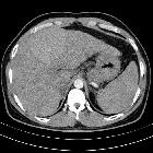
Multiple
regenerative liver nodules uncovering a constrictive pericarditis. A solid nodule at segment III is shown, almost isoechogenic to the remaining hepatic parenchyma. Several similar nodules were also found.

Multiple
regenerative liver nodules uncovering a constrictive pericarditis. T1 out-of-phase, showing a smaller nodule at segment IVa, with similar features to the one in Fig. 4a.

Multiple
regenerative liver nodules uncovering a constrictive pericarditis. The nodule in segment III is isointense on T2 without fat suppression.

Multiple
regenerative liver nodules uncovering a constrictive pericarditis. The nodule in segment III is isointense on T2 SPAIR.

Multiple
regenerative liver nodules uncovering a constrictive pericarditis. The nodule in segment IVa is isointense on T2 SPAIR.

Multiple
regenerative liver nodules uncovering a constrictive pericarditis. The nodule at segment III is isointense on T1 GRE fat sat (THRIVE) without contrast.

Multiple
regenerative liver nodules uncovering a constrictive pericarditis. The nodule in segment III is isointense on T1 GRE fat sat (THRIVE), hepatobiliary phase.

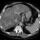
Multiple
regenerative liver nodules uncovering a constrictive pericarditis. T1 GRE fat sat (THRIVE) after Gd EOB-DTPA, arterial phase, depicting mottled pattern of enhancement of the liver parenchyma and periportal hypointensity in keeping with perivascular lymphoedema.
Regenerative
liver nodule • Macroregenerative siderotic hepatic nodule - Ganzer Fall bei Radiopaedia

Qualitative
analysis of small (≤2 cm) regenerative nodules, dysplastic nodules and well-differentiated HCCs with gadoxetic acid MRI. MR scans in a patient with chronic hepatitis, HCV related, and a regenerative nodule in liver segment VIII. a-b T1 - and T2-weighted images do not reveal any focal liver lesion. c T1-weighted gradient-echo shows a hypervascular lesion without wash-out during the late dynamic phase d). On the corresponding MR image obtained during the liver-specific hepatobiliary phase (e), the lesion is isointense to adjacent hepatic parenchyma. At pathologic examination of the explanted liver, this lesion corresponded to a multiacinar cirrhotic nodule

A small
hepatic nodule ( ≤2 cm) in cirrhotic liver: doTriphasic MRI and Diffusion-weighted image help in diagnosis. A male patient aged 63 years old presented with tender abdomen who underwent ultrasonography and revealed several small nodules and referred to the MRI unit for further evaluation. a An axial unenhanced T1WI shows multiple nodules of isointense signal similar to liver parenchyma. b An axial T2WI shows the lesion to be of low SI. c An axial post-contrast arterial phase image shows the lesion to be isointense. d, e An axial portal and delayed post-contrast image shows the lesions were also isointense with no contrast uptake. f DWI shows the lesions were isointense owing to free diffusion. Suggested MRI diagnosis: multiple regeneration nodules. Biopsy results: confirmed regeneration nodules

MR imaging in
liver cirrhosis: classical and new approaches. Fibrosis and regenerative nodules. Transverse STIR image shows a reticular pattern of the fibrotic bands surrounding hypointense regenerative nodules

MR imaging in
liver cirrhosis: classical and new approaches. Non-tumoral nodules. Transverse opposed (a) and in-phase (b) T1W images show hyperintense nodules without decreased signal intensity on opposed-phase imaging and hypointensity on STIR image (c). Note several Gamna-Gandy siderotic splenic bodies due to advanced portal hypertension

MR imaging in
liver cirrhosis: classical and new approaches. Fibrosis. Transverse fat suppression T1W spoiled GRE image after administration of contrast media shows reticular pattern of the fibrotic bands surrounding regenerative nodules
 nicht verwechseln mit: noduläre regenerative Hyperplasie
Regeneratknoten der Leber
Siehe auch:
- Leberzirrhose
- noduläre regenerative Hyperplasie
- adenomatöse Hyperplasie Leber
- dysplastische Leberknoten
und weiter:


 Assoziationen und Differentialdiagnosen zu Regeneratknoten der Leber: