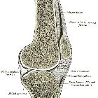
Hoffa-Fettkörper


Knee bursae
• Knee anatomy (illustrations) - Ganzer Fall bei Radiopaedia

Infrapatellar
fat pad • Hoffa's fat pad (Gray's illustration) - Ganzer Fall bei Radiopaedia

Infrapatellar
fat pad • Normal knee fat pads in the anterior knee compartment - Ganzer Fall bei Radiopaedia

Hoffa’s fat
pad abnormalities, knee pain and magnetic resonance imaging in daily practice. Anatomy of the HFP. The HFP (Hoffa fp) is limited anteriorly by the patellar tendon (Pat ten) and the joint capsule, superiorly by the inferior pole of the patella (Pat) (a), inferiorly by the proximal tibia (Tib) and the deep infrapatellar bursa (asterisk), and posteriorly by the synovium (arrows) and femur (Fem). It is attached directly to the anterior horns of the menisci (Med men, Lat men) (b). Normal vascular supply consists of two vertical arteries, posterior and parallel to the lateral edges of the patellar tendon (c)
The infrapatellar fat pad, also known as Hoffa fat pad, is the largest of the anterior knee fat pads. It is located immediately posterior to the patellar tendon and is traversed during knee arthroscopy.
Anatomy
Boundaries
- anterior
- anteroinferior: patellar tendon
- anterosuperior: patella
- posterior
- posterocentral: knee joint (femorotibial articulation)
- posteroinferior: tibia
- posterosuperior: femur
Related pathology
- patellar tendon lateral femoral condyle friction syndrome (Hoffa fat pad impingement syndrome)
- ganglion cyst of Hoffa fat pad
Siehe auch:
und weiter:

 Assoziationen und Differentialdiagnosen zu Hoffa-Fettkörper:

