abdominale Lymphangiomatose

Abdominal and
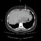
thoracic lymphangioma with unusual imaging. Diffuse tissue thickening is demonstrated around the liver (white arrow) containing calcification (red arrow).

Abdominal and
thoracic lymphangioma with unusual imaging. Coronal T1 MRI pre-contrast with fat -aturation shows diffuse hypointensity around the liver corresponding to the finding on CT (arrows).

Abdominal and
thoracic lymphangioma with unusual imaging. Coronal T1 MRI post-contrast shows minimal patchy enhancement of the lesion.

Abdominal and
thoracic lymphangioma with unusual imaging. Coronal MRI T2 Haste-fs shows the hyperintense perihepatic masses with septations (arrow).

Abdominal and
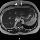
thoracic lymphangioma with unusual imaging. Axial MRI T2 Haste shows calcification (long arrow) and septae (short arrow).

Abdominal and
thoracic lymphangioma with unusual imaging. This shows the density of the surrounding soft tissue (40 HU).

Abdominal and
thoracic lymphangioma with unusual imaging. Coronal CT post-contrast porto-venous phase demonstrates the abnormal tissue thickening around the liver and the hepatorenal recess.
abdominale Lymphangiomatose
Von einer Lymphangiomatose spricht man, wenn nicht ein einzelnes Lymphangiom als singuläre Raumforderung vorliegt, sondern wenn die Fehlbildungen weitverbreitet oder an verschiedenen Lokalisationen im Körper vorkommen.
Siehe auch:

 Assoziationen und Differentialdiagnosen zu abdominale Lymphangiomatose:

