bihiläre Lymphadenopathie

School ager
with a cough. CXR PA and lateral shows massive bilateral hilar lymphadenopathy with the lungs being clear.The diagnosis was tuberculosis.


Sarkoidose in
der Computertomographie: Querschnitt durch den Thorax im Bereich der Aufzweigungen der Bronchien (der Hili) mit vielen vergrößerten Lymphknoten (Pfeile).

Pulmonary
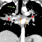
sarcoidosis. Conglomerate bilateral perihilar masses are seen extending along peribronchovascular interstitium radiating to periphery (star). Note enlarged subcarinal (orange arrow) and bilateral hilar regions (yellow arrows).

Pulmonary
sarcoidosis. Conglomerate bilateral perihilar masses are seen extending along peribronchovascular interstitium radiating to periphery (star). Note enlarged right paratracheal (green arrows), subcarinal (orange arrow) and bilateral hilar regions (yellow arrows)

Pulmonary
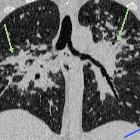
sarcoidosis. Conglomerate perihilar masses (star) extending along the peribronchovascular interstitium. Note the bronchi traversing through these masses without their obliteration. Mulitple well-defined nodules are seen along the peribronchovascular interstitium (green arrows) and subpleural areas (blue arrow).

Pulmonary
sarcoidosis. Mulitple well-defined nodules are seen along the peribronchovascular interstitium (green arrows) and subpleural areas (blue arrow).
bihiläre Lymphadenopathie
Siehe auch:

 Assoziationen und Differentialdiagnosen zu bihiläre Lymphadenopathie:


