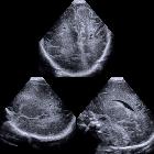
konnatale Zysten neben den Ventrikelvorderhörnern

Intracerebral
periventricular cysts in newborn: Connatal cysts. Illustration depicting the various locations of intracerebral cystic anomalies encountered in the newborn. CC= Connatal cyst SC= Subependymal cyst PVL= Periventricular Leukomalacia

Premature
newborn screening head US exam. Coronal (above) and sagittal (below) US of the brain show bilaterally multiple small cysts just lateral to the superolateral angles of the anterior horns of the lateral ventricles.The diagnosis was connatal cyst.

Connatal cyst
• Connatal cysts - Ganzer Fall bei Radiopaedia

Connatal cyst
• Connatal cysts - Ganzer Fall bei Radiopaedia

Connatal cyst
• Connatal cyst - Ganzer Fall bei Radiopaedia

Intracerebral
periventricular cysts in newborn: Connatal cysts. Transfontanellar coronal ultrasound image of the brain reveals simple, oblong cysts adjacent and along the supero-lateral margins of both frontal horns.

Intracerebral
periventricular cysts in newborn: Connatal cysts. Transfontanellar sagittal ultrasound image of the brain reveals simple, oblong cysts adjacent to the anterior aspect of the brain ventricles.
Connatal cysts, also known as coarctation of the lateral ventricles or frontal horn cysts, are cystic areas adjacent to the superolateral margins of the body and frontal horns of the lateral ventricles and are believed to represent a normal variant.
Epidemiology
The incidence is 0.7% in low birth weight preterm infants.
Pathology
It was previously believed to be a sequela of different insults in the developing brain , however, it seems to be just a normal variant . The outward most part of the ventricle attains a rounded appearance when the ventricle walls are close enough and touching each other. This gives a cystic appearance sonographically.
Radiographic features
Ultrasound
- thin walled cysts in the superior and lateral aspect of frontal horn of lateral ventricle and anterior to the foramina of Monro
- most often multiple cysts noted
- a string of pearls appearance may be seen
- regresses on follow up scan
Treatment and prognosis
They have been shown to regress on follow-up imaging.
Differential diagnosis
Considerations include:
See also
Siehe auch:
- periventrikuläre Leukomalazie
- Entwicklungsstörungen des Ventrikelsystems
- fetal intracranial cystic lesions
- pseudocysts of the germinal matrix
- Entwicklungsstörung Seitenventrikel
- subependymale Zyste
- periventrikuläre Arachnoidalzysten
und weiter:

 Assoziationen und Differentialdiagnosen zu konnatale Zysten neben den Ventrikelvorderhörnern:
pseudocysts
of the germinal matrix


