verdickte Magenwand

B-Zell-Lymphom
des Magens mit erheblicher Verdickung der Magenwand, wobei der Tumor eine relativ homogene Dichte aufweist. Kleinkurvaturseitig mehrere Lymphknoten auch paragastral.

Phlegmonöse
Gastritis in der Computertomographie: Die diffuse, den ganzen Magen betreffende hypodense Wandverdickung trat innerhalb eines Tages nach einer Endoskopie mit Probenentnahme auf.

Phlegmonöse
Gastritis in der Computertomographie: Die diffuse, den ganzen Magen betreffende hypodense Wandverdickung trat innerhalb eines Tages nach einer Endoskopie mit Probenentnahme auf.

Phlegmonöse
Gastritis in der Computertomographie: Die diffuse, den ganzen Magen betreffende hypodense Wandverdickung trat innerhalb eines Tages nach einer Endoskopie mit Probenentnahme auf.
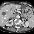
Riesenfaltengastritis
in der Computertomographie axial. Mit abgebildete Leberzysten.
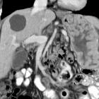
Riesenfaltengastritis
in der Computertomographie coronar rekonstruiert. Mit abgebildete Leberzysten.

Linitis
plastica • Gastric Burkitt lymphoma - Ganzer Fall bei Radiopaedia

Gastric
lymphoma • Gastric lymphoma - Ganzer Fall bei Radiopaedia

Gastric wall
fatty infiltration • Gastric wall fatty infiltration - Ganzer Fall bei Radiopaedia

Gastric
lipomatosis treated by total gastrectomy: a case report. Findings on EUS and CT. EUS (a) and CT (b, c) findings are shown. EUS shows a high-echoic lesion in the antral wall submucosa (red triangle), extending to the stomach. CT shows a huge fat-containing mass lesion around the gastric wall, excluding the lesser curvature (red triangle). Abbreviations: CT computed tomography, EUS endoscopic ultrasound

Gastric
lipomatosis treated by total gastrectomy: a case report. Findings on T1- and T2-weighted MRI. T1- (a) and T2-weighted (b) MRI findings are shown. There is a high-intensity lesion in the submucosa with a fat-containing mass on the entire wall of the gastric antrum and body, but excluding the lesser curvature (red triangle). Abbreviations: MRI magnetic resonance imaging


 Assoziationen und Differentialdiagnosen zu verdickte Magenwand:

