genitourinary tuberculosis

Extrapulmonary
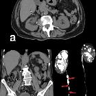
tuberculosıs: an old but resurgent problem. Axial (a) and coronal (b) non-contrast CT scan of a 74-year-old male with urinary tuberculous reveal debris collection within the dilated right renal pelvis and dilated right ureter with diffuse wall thickening. 3D MIP Coronal T2-weighted MR urogram shows strictures (arrows) of the right ureter

Extrapulmonary
tuberculosıs: an old but resurgent problem. A 47-year-old male. Coronal post-contrast CT scan shows diffuse thickening of the bladder wall (arrow) and dilated left proximal ureter (arrowheads). Urinary TB

Urinary
tuberculosis: multidetector CT findings. Nephrographic acquisition (b...f) showed left kidney with normal size and function, mild hydronephrosis (*). Right-sided findings included decreased renal parenchyma and delayed enhancement, uneven caliectasis (thin arrows), mild enhancing urothelial thickening (thick arrows).

Genitourinary
tuberculosis • Disseminated tuberculosis to wrist and testes - Ganzer Fall bei Radiopaedia

Differential
diagnosis of uncommon prostate diseases: combining mpMRI and clinical information. Genitourinary tuberculosis in a 26-year-old man with a right scrotal skin ulceration accompanied by pus discharge and a strongly positive tuberculin skin test. a Computed tomography image shows an enhanced nodule in the right epididymis (arrowhead) with testicular hydrocele (arrow). b T2WI shows prostate atrophy and multiple extremely hypointense nodules inside the tissue (arrowheads). c T2WI shows slightly inhomogeneous signal intensity of the prostate. d DWI at a b-value of 800 s/mm2 shows slight hypointensity of the nodules (arrowheads)
Genitourinary tuberculosis is the second most common site of infection in humans by Mycobacterium tuberculosis, second only to pulmonary tuberculosis.
It can most easily be divided anatomically into:
- renal tuberculosis (renal parenchyma, calyces and renal pelvis)
- bladder and ureteric tuberculosis
- prostatic tuberculosis
- scrotal tuberculosis (testes, epididymis, seminal vesicles, ductus deferens)
- tuberculous pelvic inflammatory disease (female)
For a general discussion of the disease, refer to the article on tuberculosis.
Siehe auch:
und weiter:

 Assoziationen und Differentialdiagnosen zu urogenitale Tuberkulose:


