parotid space
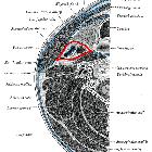
The parotid space is one of the deep compartments of the head and neck and, as the name suggests, is mostly filled with the parotid gland. It is the most lateral major suprahyoid neck space.
Gross anatomy
The parotid space is a roughly pyramidal space, the broad elongated base facing laterally, formed by the superficial layer of the deep cervical fascia overlying the superficial lobe of the parotid gland, and its apex pointing medially. It is traversed by the external carotid artery, retromandibular vein and facial nerve.
Contents
- parotid glands
- intraparotid lymph nodes
- intraparotid facial nerve (CN VII)
- external carotid artery
- retromandibular vein
Relations
- lateral to the parapharyngeal space
- medial to superficial space and subcutaneous tissue
- anterior to the carotid space
- posterior belly of the digastric muscle forms a variable portion of the posteromedial border of the parotid space and at times this muscular band helps to differentiate a deep-lobe parotid space lesion to one arising in the carotid space
- posterior to the masticator space
Boundaries
The parotid space is circumscribed by the superficial layer of the deep cervical fascia :
- superior margin: external auditory canal; apex of the mastoid process
- inferior margin: inferior mandibular margin (although the parotid tail can extend further inferiorly below the angle of the mandible)
- anterior margin: masticator space
Radiographic appearance
Ultrasound
- with the parotid gland filling around two-thirds of the parotid space, US findings will reflect those of the parotid gland
- intraparotid lymph nodes (may number up into the twenties) are also evident as rounded/bean-shaped hypoechoic structures with an echogenic central fatty hilum within the hyperechoic parotid gland
- most frequently located in the preauricular region and they provide lymphatic drainage of the external ear and lateral scalp
- external carotid artery and retromandibular vein will be found posteriorly
- retromandibular vein crosses the superficial lobe of the parotid gland and continues longitudinally until it reaches the inferior margin of the parotid gland to join with the external jugular vein
- external carotid artery travels in the same route, however, it is a larger structure and is found in a deeper plane to the retromandibular vein
- facial nerve is not visualized on US, but is inferred to be located lateral to the retromandibular vein
CT
- lower attenuation in comparison to muscle due to the combination of fat and glandular tissue
- allows for good visualization of deep lesions as well as reliable assessment of size, location, margins, extracapsular extension, involvement of adjacent structures, and areas of necrosis, hemorrhage, calcification of cysts
- CT is preferred for tender, recurrent parotid masses that are likely to be inflammatory whereas MRI is better for assessment of a painless parotid lump
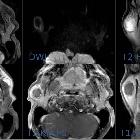
MRI
- T1: higher signal than muscle
- T2: lower signal than muscle
- MRI is preferred for imaging the parotid space in children
- neonates and young children have limited amounts of fat within the parotid gland which decreases the amount of natural contrast available on CT, which increases the difficulty to perceive a mass lesion and define its margins.
- MRI is significantly less affected by the relative lack of native fat
Related pathology
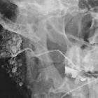
Any mass originating from the parotid space will be centered within the parotid gland.
A large mass arising within the deep lobe of the parotid will medially displace fat in the parapharyngeal space and cause posteromedial displacement of the posterior belly of the digastric muscle and carotid space. There is also often associated widening of the stylomandibular notch .
- congenital
- salivary gland tumors
- benign tumor
- primary malignant tumor
- metastatic malignant tumor
- metastatic adenopathy
- lymphoma
- parotid cysts
- lipoma
- inflammatory
- sialadenitis
- chronic granulomatous parotitis
- abscess/cellulitis
- Sjogren syndrome/autoimmune
- benign lymphoepithelial cysts (AIDS)
- nodular fasciitis
- reactive adenopathy
Siehe auch:
- Arteria carotis externa
- carotid space
- Glandula parotidea
- Tumoren der Speicheldrüsen
- deep compartments of the head and neck
- masticator space
- zystische Läsionen der Glandula parotis
- Retropharyngealraum
- superficial mucosal space
- prevertebral space
- Parapharyngealraum
und weiter:

 Assoziationen und Differentialdiagnosen zu parotid space:


