colloid cyst

Zufallsbefund
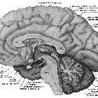
einer Kolloidzyste am Foramen Monroi. Links sagittale Magnetresonanztomographie, rechts zur Korrelation anatomisches Bild von Gray (File:Gray715.png). Der Befund wurde nicht histologisch gesichert, ist aber typisch.

Zufallsbefund
einer Kolloidzyste am Foramen Monroi. Links axiale native Computertomographie, rechts coronare ebenfalls native MRT. Der Befund wurde nicht histologisch gesichert, ist aber typisch.

Zufallsbefund
einer Kolloidzyste am Foramen Monroi. Magnetresonanztomographie: Links T1 coronar nativ, rechts T2 sagittal. Der Befund wurde nicht histologisch gesichert, ist aber typisch.

Zufallsbefund
einer Kolloidzyste am Foramen Monroi. Links axiale native Computertomographie, rechts coronar. Der Befund wurde nicht histologisch gesichert, ist aber typisch.

Symptomatic
colloid cyst. Plain CT transversal showing the hyperdense colloid cyst.

Symptomatic
colloid cyst. MRI FLAIR transversal showing the hyperintense intraventricular colloid cyst.

Symptomatic
colloid cyst. MRI T2 coronal showing the intraventricular colloid cyst being hypointense compared to the cerebrospinal fluid.

Symptomatic
colloid cyst. MRI T1 transversal. The lesion is isointense to the cerebrum.

Symptomatic
colloid cyst. MRI T1 post-contrast (7 ml Gadovist) – no enhancement of the lesion.

 Assoziationen und Differentialdiagnosen zu Kolloidzyste:
