Scleroderma (musculoskeletal manifestations)

Erosive
Arthropathy in systemic sclerosis. X-ray of hands showing. - Erosion of the fourth and the fifth proximal inter phalangeal joint on the left hand. - Terminal phalangeal tuft resorption (arrow).

Erosive
Arthropathy in systemic sclerosis. X-ray of hand showing: a small and discrete lesion in the fourth metacarpo-phalangeal joint similar to early rheumatoïd arthritis.

Erosive
Arthropathy in systemic sclerosis. X-ray of hands showing a joint space narrowing in the proximal inter phalangeal joint similar to erosive osteoarthritis.

Erosive
Arthropathy in systemic sclerosis. X-ray of a left foot. Erosive lesion in the first metatarso-phalangeal joint, and subluxation and erosion of the second proximal inter phalangeal joint similar to psoriatic arthritis.

Scleroderma
• Scleroderma - hand manifestations - Ganzer Fall bei Radiopaedia

Scleroderma
• Scleroderma - Ganzer Fall bei Radiopaedia

Scleroderma
(musculoskeletal manifestations) • Scleroderma - Ganzer Fall bei Radiopaedia

Scleroderma
(musculoskeletal manifestations) • Scleroderma - hand manifestations - Ganzer Fall bei Radiopaedia

Scleroderma
• Progressive systemic sclerosis involving the knee - Ganzer Fall bei Radiopaedia

Scleroderma
(musculoskeletal manifestations) • Scleroderma - hand manifestations - Ganzer Fall bei Radiopaedia

Scleroderma
(musculoskeletal manifestations) • Systemic sclerosis - Ganzer Fall bei Radiopaedia

Scleroderma
(musculoskeletal manifestations) • Scleroderma - Ganzer Fall bei Radiopaedia

Scleroderma
(musculoskeletal manifestations) • Scleroderma - Ganzer Fall bei Radiopaedia

Scleroderma
(musculoskeletal manifestations) • Scleroderma - Ganzer Fall bei Radiopaedia
Musculoskeletal manifestations of scleroderma are common and variable.
For a general discussion of scleroderma, please refer to the parent article: scleroderma.
Radiographic features
Plain radiograph
Imaging findings demonstrate bone and soft tissue changes. The hands are the most common site of involvement.
Bone changes:
- acro-osteolysis (resorption of the distal phalanges)
- periarticular osteopenia
- joint space narrowing
- erosions
- severe resorption of the first CMC joint with radial subluxation is a characteristic feature on hands radiographs
Soft tissue changes:
- subcutaneous and periarticular calcification
- atrophy especially at tips of fingers
- flexion contractures
Other less common documented musculoskeletal findings:
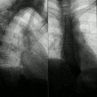
- rib resorption (bilateral superior rib notching, predominantly along posterior surface), mandibular angle resorption (+/- loss of lamina dura), radius and ulna resorption
- terminal phalangeal sclerosis
Differential diagnosis
See also
- pulmonary manifestations of scleroderma
- cardiac manifestations of scleroderma
- gastrointestinal manifestations of scleroderma
- hepatobiliary manifestations of scleroderma
- renal manifestations of scleroderma
- differential diagnosis of erosive arthritis
Siehe auch:
- Systemische Sklerodermie
- Akroosteolyse
- pulmonary manifestations of scleroderma
- gastrointestinale Manifestationen Sklerodermie
- Arthropathie
und weiter:


 Assoziationen und Differentialdiagnosen zu Systemic sclerosis (musculoskeletal manifestations):