Benign esophageal lesions

Leiomyome des
Ösophagus: Links und Mitte: Fälle aus der Breischluckuntersuchung: Glatte Begrenzung der in das Lumen ragenden Raumforderung. Oberhalb ist das Lumen durch die peristaltische Kontraktion nicht mehr sichtbar (links). Der Winkel zur angrenzenden Lumenkontur ist stumpf (submuköse Lage). Rechts: In der CT kann die rundliche Konfiguration des Tumors oft erst in sagittalen oder koronaren Reformatierungen dargestellt werden.
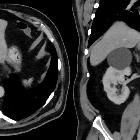
Submuköses
Lipom im unteren Ösophagus bei einem Mann. Computertomographie axial und coronar.

Benign
esophageal lesions • Esophageal duplication cyst - Ganzer Fall bei Radiopaedia


Fibrovaskulärer
Polyp in der Schluckuntersuchung: (a) Der intraluminale Tumor dehnt sich nahezu über die ganze Länge der Speiseröhre aus. (b) Als Reaktion auf den chronischen Fremdkörperreiz lassen sich zirkuläre oder spiralige Kontraktionen beobachten.


Oesophageal
schwannoma. CT with sagittal view showing a low-density mass surrounded by the trachea and vertebrae. The tumour also compresses the trachea.

Successful
removal of a giant esophageal lipoma by thoracoscopic enucleation: a case report. Preoperative imaging. (a horizontal, b coronal) CT scan of the chest revealed a 10 × 7 cm homogenous mass in the middle and lower esophagus. c Upper endoscopy revealed a submucosal tumor with normal mucosa arising from the left esophageal wall

Video-thoracoscopic
enucleation of esophageal leiomyoma. A. Esophagogram of Case 10. Reveals typical findings of intramural tumor (T) near the esophago-gastric junction: smooth surface, clear-cut margins, and sharp angles at upper and lower ends of the tumor. E: esophagus. B. Endoscopic view of the esophageal leiomyoma (T). (Case 10).

Video-thoracoscopic
enucleation of esophageal leiomyoma. Computed tomography, cross section (A). Reveals the esophageal lumen being compressed forwards by horse-shoe shaped (T1 and T2) leiomyoma. Coronal section (B). Reveals this tumor (T) compressing the esophagus (E) and adjacent structures of mediastinum. (Case 9).

Imaging of
the oesophagus: beyond cancer. Hemangioma. CT angiogram of the chest in soft tissue window shows a round calcification within the wall of the oesophagus compatible with a phlebolith. This finding is most often seen in patients with varices, however this patient was found to have a hemangioma

Resection of
an esophageal schwannoma with thoracoscopic surgery: a case report. CT and FDG-PET CT findings. a Computed tomography revealed a large isodense mass of the esophageal wall in the upper mediastinal space. b Accumulation of fluorodeoxyglucose is demonstrated in the upper thoracic esophagus

Esophageal
leiomyoma • Esophageal leiomyoma - Ganzer Fall bei Radiopaedia

Imaging of
the oesophagus: beyond cancer. Leiomyoma. Esophagram in AP projection demonstrates a smooth, lobulated filling defect within the mid-thoracic oesophagus at the level of the carina. Notice the filling defect makes an acute angle to the oesophageal wall, suggesting that this lesion is intrinsic to the oesophagus
Benign esophageal lesions are less symptomatic than malignant esophageal lesions, making up for only 1% of clinically apparent esophageal lesions.
Pathology
- esophageal leiomyoma (>50%)
- may calcify
- esophageal fibrovascular polyp (~12.5%)
- may contain fat
- esophageal duplication cyst (10%)
- oesphageal papilloma (3%)
- oeseophageal fibroma (3%)
- esophageal hemangioma (2%)
- esophageal hamartoma
- esophageal neurofibroma
Siehe auch:
- Hämangiom
- Leiomyom Ösophagus
- Ösophagusstrikur
- Fibrovaskulärer Polyp des Ösophagus
- maligne Ösophagustumoren
- Granularzelltumor des Ösophagus
- Lipome des Ösophagus
- Neurofibrom des Ösophagus
- Leiomyomatose des Ösophagus
- Hämangiom des Ösophagus
- Schwannom des Ösophagus
- inflammatory fibroid polyp of the oesophagus
- papilloma of the esophagus
- oseophageal cancer
und weiter:


 Assoziationen und Differentialdiagnosen zu benigne Ösophagustumoren:


