patellar dislocation


Lateral patellar dislocation refers to lateral displacement followed by dislocation of the patella due to disruptive changes to the medial patellar retinaculum.
Epidemiology
Patellar dislocation accounts for ~3% of all knee injuries and is commonly seen in those individuals who participate in sports activities.
Pathology
Patellar dislocation most commonly results from a twisting motion, with the knee in flexion and the femur rotating internally on a fixed foot (valgus-flexion-external rotation) .
Radiographic features
Plain radiograph
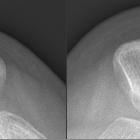
- lateral displacement of patella noted on skyline projection
- joint effusion
- sliver sign
MRI
The following features are noted:
- medial retinacular abnormalities (ranging from strain to complete disruption) with adjacent periligamentous edema and hemorrhage
- lateral displacement of patella (not necessarily seen in transient dislocation)
- medial patellar contusion +/- corresponding lateral femoral condyle contusion
- joint effusion
The presence of an abnormal medial patellar retinaculum should suggest the diagnosis of transient lateral patellar dislocation .
The images should be scrutinized for the presence of chondral or osteochondral injury, especially if displaced as an intra-articular body, as this may affect surgical management.
The trochlear groove and patella may have abnormal morphology that predisposes to patellar dislocation.
Differential diagnosis
- acute ACL tear: no medial patellar contusion in this injury
- direct trauma to lateral knee: normally no patellar contusion
Siehe auch:
- Jägerhutpatella
- Patelladysplasie
- mediales Retinaculum patellae
- Operation nach Blauth (Patellasehne)
- Scholte-Syndrom
- patella fracture-dislocation
- Ruptur mediales Retinaculum
- Patellaluxation bei Trisomie 18
- patellar subluxation
- Roux-Goldthwait procedure
- Prieto-Syndrom
- chronische Patellaluxation
und weiter:

 Assoziationen und Differentialdiagnosen zu Patellaluxation:

