anterior cruciate ligament tear


Anterior cruciate ligament (ACL) tears are the most common knee ligament injury encountered in radiology and orthopedic practice.
Clinical presentation
Patients typically present with symptoms of knee instability, usually after acute trauma. The following signs and symptoms are common:
- popping sensation at the time of injury, followed by swelling
- initial inability to weight wear, which improves in a short period
- knee felt to "gives way" especially during pivoting movement
- apprehension with an attempt at non-linear movements
The combination of the Lachman, pivot shift and anterior drawer tests are used to clinically confirm diagnosis .
Pathology
The anterior cruciate ligament is the most commonly disrupted ligament of the knee, especially in athletes who participate in sports that involve rapid starting, stopping, and pivoting (e.g. soccer, basketball, tennis, netball, and snow skiing).
Associations
- O'Donoghue's unhappy triad
- Segond fracture
- posteromedial corner injury of the knee
- Meniscocapsular separation
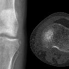
Radiographic features
In younger patients, avulsion of the tibial attachment may be seen.
Plain radiograph
- deep lateral sulcus sign - depression of lateral femoral condyle representing impaction fracture
- anterior tibial translocation sign
- Segond fracture
- arcuate fracture
- joint effusion
CT
Considered to have high specificity and sensitivity in detecting anterior cruciate ligament disruption . CT is helpful in characterizing the avulsion bone fragment when it is present.
MRI
Imaging of anterior cruciate ligament tears should be divided into primary and secondary signs.
Primary signs are those that pertain to the ligament itself. Secondary signs are those which are closely related to anterior cruciate ligament injuries.
Primary signs
- swelling
- increased signal on T2 or fat-saturated PD
- fiber discontinuity
- abnormal anterior cruciate ligament orientation relative to intercondylar (Blumensaat's) line
- ACL fibers subjectively less steep than a line tangent to the intercondylar roof (Blumensaat's line)
- ACL angle (angle between the intercondylar line and ACL) >15° with the apex of the angle located anteriorly, indicating a less steep ACL line - this indicates a ruptured and collapsed ligament
- empty notch sign: a fluid signal at the site of femoral attachment at the intercondylar notch, denotes avulsion at the femoral attachment.
ACL tears typically occur in the middle portion of the ligament (midsubstance tears) and appear as discontinuity of the ligament or abnormal contour. The signal of the ACL can be more hyperintense on T2. If the angle is still normal and there is a hyperintense signal, a partial rupture is more likely than a complete rupture.
ACL tear may only involve one bundle. Imaging signs of isolated posterolateral bundle tear are as follows:
- gap sign: fluid signal and/or a gap between the medial aspect of the lateral femoral condyle and the lateral aspect of the mid-ACL, can be seen on either axial or coronal MRI images.
- footprint sign: incomplete coverage of the lateral aspect of the tibial spine of the tibia by the distal ACL attachment, seen only on coronal MRI images
Secondary signs
Secondary signs include :
- bone contusion in lateral femoral condyle and posterolateral tibial plateau
- >7 mm of anterior tibial translation, also known as the anterior tibial translocation sign or anterior drawer sign
- uncovered posterior horn of the lateral meniscus
- Segond fracture, and to a lesser degree arcuate sign
- reduced PCL angle due to buckling of PCL
- positive PCL line sign
- medial or lateral collateral ligament injury
Related video
Treatment and prognosis
Anterior cruciate ligament reconstruction aims to reduce joint instability and avoid (further) meniscal and/or cartilage damage. However, ~17.5% (range 13.6-21.5%) of patients develop symptomatic osteoarthritis post ACL reconstruction .
See also
Siehe auch:
- Segond-Fraktur
- deep lateral femoral notch (sulcus) sign
- unhappy triad
- arcuate sign
- vordere Kreuzbandplastik
- Teilruptur vorderes Kreuzband
- Komplikationen nach vorderer Kreuzbandplastik
- anterior tibial translocation sign
und weiter:
- mukoide Degeneration des vorderen Kreuzbandes
- Endobutton
- Contre-coup-Verletzung am Kniegelenk
- Kreuzband Signalsteigerung
- hintere Kreuzbandruptur
- Verletzungen hinteres Kreuzband
- meniscal tear patterns in ACL disruption
- spontaneous healing of complete ACL tear
- O'Donoghue's unhappy triad
- Kreuzbandriss
- vorderes Kreuzband des Kniegelenks
- Anterolaterales Ligament
- anterior cruciate ligament bony avulsion
- Pivot-Shift-Test
- ACL graft tear
- posterolateral corner injury


 Assoziationen und Differentialdiagnosen zu Ruptur des vorderen Kreuzbandes: