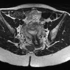
arcuate uterus


An arcuate uterus is a mildly variant shape of the uterus. It is technically one of the Müllerian duct anomalies, but is often classified as a normal variant. It is the uterine anomaly that is least commonly associated with reproductive failure. Arcuate uterus can be characterized with ultrasound or MRI.
Pathology
An arcuate uterus is characterized by a mild indentation of the endometrium at the uterine fundus. It occurs due to a failure of complete resorption of the uterovaginal septum, and is the most common Mullerian duct anomaly, affecting 3.9% of the general population .
Radiographic features
General features include:
- normal fundal contour
- no division of uterine horns
- smooth indentation of fundal endometrial canal: the depth of indentation is usually considered to be <1 cm
- increased transverse diameter of uterine cavity
Fluoroscopy
On hysterosalpingograms there is opacification of the endometrial cavity demonstrates a single uterine canal with a broad saddle-shaped indentation of the uterine fundus.
Pelvic ultrasound
A normal external uterine contour is noted, with a broad smooth indentation on the fundal segment of the endometrium. There should not be division of the uterine horns.
MRI
A normal external uterine contour is maintained. The myometrial fundal indentation is smooth and broad, and the signal intensity of this region is isointense to normal myometrium.
Differential diagnosis
- septate uterus
- arcuate uterus and septate uterus exist on a spectrum from most to least resorption of the uterovaginal septum, respectively
- bicornuate uterus
- arcuate uterus can be distinguished from a bicornuate uterus on the basis of its complete fundal unification (i.e. an arcuate uterus has a normal or slightly indented external fundal contour, whereas a bicornuate uterus has a more marked fundal indentation, ≤5 mm above the level of the uterine horns)
Siehe auch:
und weiter:

 Assoziationen und Differentialdiagnosen zu Uterus arcuatus:

