articular ecchondrosis

Synovialchondromatose
des Hüftgelenks links. Histologisch nicht gesichert, jedoch radiologisch typischer Befund.

Case report:
Primary synovial chondromatosis of the right TMJ.. Coronal and sagittal CT (bone window) show multiple calcified loose bodies of uniform size and widening of the right TMJ (red circle).

Synovial
chondromatosis • Synovial chondromatosis - Ganzer Fall bei Radiopaedia

Synoviale
Chondromatose mit multiplen Chondromen in einer Bakerzyste bei Gonarthrose

Synovial
chondromatosis • Synovial osteochondromatosis (illustration) - Ganzer Fall bei Radiopaedia

Radiological
identification and analysis of soft tissue musculoskeletal calcifications. Synovial osteochondromatosis. Anteroposterior radiograph of the left hip shows multiple intra-articular osteocartilaginous bodies (arrows) with ring-like calcifications centred on the coxofemoral joint


Synovialchondromatose
Schulter: Verteilung auch bis in die verschiedenen Gelenkausläufer.

Primary
synovial chondromatosis • Synovial osteochondromatosis - Ganzer Fall bei Radiopaedia

Apple core
sign (femur) • Synovial chondromatosis - Ganzer Fall bei Radiopaedia

Synovialchondrome
in Bakerzyste bei Gonarthrose. Mit im Bild eine Kalibrierungskugel für die Vorbereitung zur Prothesenimplantation.

Synovial
chondromatosis • Synovial osteochondromatosis - Ganzer Fall bei Radiopaedia

Synovial
chondromatosis • Primary synovial osteochondromatosis - Ganzer Fall bei Radiopaedia

Synovial
chondromatosis • Synovial chondromatosis - Ganzer Fall bei Radiopaedia

Primary
synovial chondromatosis • Ulnocarpal synovial osteochondromatosis - Ganzer Fall bei Radiopaedia

Synovial
chondromatosis • Synovial chondromatosis - knee - Ganzer Fall bei Radiopaedia
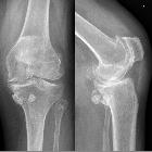
Synovialchondromatose
Kniegelenk: dorsomedial, somit am ehesten in einer Baker-Zyste.

Synovial
chondromatosis • Synovial chondromatosis - hand - Ganzer Fall bei Radiopaedia

Synovial
chondromatosis • Synovial chondromatosis of knee - Ganzer Fall bei Radiopaedia

Primary
synovial chondromatosis • Primary synovial chondromatosis - Ganzer Fall bei Radiopaedia

Primary
synovial chondromatosis • Synovial osteochondromatosis - temporomandibular joint - Ganzer Fall bei Radiopaedia

Primary
synovial chondromatosis • Synovial chondromatosis - presumed primary - Ganzer Fall bei Radiopaedia

Primary
synovial chondromatosis • Primary synovial chondromatosis of the temporomandibular joint - Ganzer Fall bei Radiopaedia

MRI imaging
of soft tissue tumours of the foot and ankle. Synovial chondromatosis. a Sagittal T2FS image of the ankle demonstrating a lobulated T2 hyperintense lesion with a low signal rim (arrows) and b avid synovial enhancement on post contrast T1FS imaging (arrows). c Mineralisation is better appreciated on the corresponding radiograph (arrows).

Synovial
chondromatosis • Synovial chondromatosis - Ganzer Fall bei Radiopaedia

Primary
synovial chondromatosis • Synovial chondromatosis of temporomandibular joint - Ganzer Fall bei Radiopaedia

Primary
synovial chondromatosis • Synovial osteochondromatosis - Ganzer Fall bei Radiopaedia

Primary
synovial chondromatosis • Primary synovial osteochondromatosis - Ganzer Fall bei Radiopaedia

Primary
synovial chondromatosis • Synovial osteochondromatosis of the shoulder joint - Ganzer Fall bei Radiopaedia

Synovial
chondromatosis • Synovial chondromatosis of the knee - Ganzer Fall bei Radiopaedia

Synovial
chondromatosis • Synovial osteochondromatosis - Ganzer Fall bei Radiopaedia

Synovial
chondromatosis • Synovial osteochondromatosis of the shoulder joint - Ganzer Fall bei Radiopaedia

Primary
synovial chondromatosis • Synovial osteochondromatosis - Ganzer Fall bei Radiopaedia

Primary
synovial chondromatosis • Primary synovial osteochondromatosis of the gastrocnemius semimembranosus bursa - Ganzer Fall bei Radiopaedia


Feasibility
of MRI in diagnosis and characterization of intra-articular synovial masses and mass-like lesions. Right knee synovial chondromatosis in 48-year-old male. Sagittal T1 (a), sagittal T2 (b), sagittal STIR (c), coronal STIR (d), and axial STIR (e) show non-mineralized equal size nodules showing low signal at T1 (white arrow), high at T2 and STIR (red arrows)

Primary
synovial chondromatosis • Distribution of synovial osteochondromatosis (diagram) - Ganzer Fall bei Radiopaedia

Primary
synovial chondromatosis • Osteochondromatosis - Ganzer Fall bei Radiopaedia

Primary
synovial chondromatosis • Synovial osteochondromatosis - Ganzer Fall bei Radiopaedia

Primary
synovial chondromatosis • Primary synovial chondromatosis - Ganzer Fall bei Radiopaedia

Primary
synovial chondromatosis • Synovial osteochondromatosis - hip - Ganzer Fall bei Radiopaedia

Primary
synovial chondromatosis • Synovial osteochondromatosis - Ganzer Fall bei Radiopaedia

Primary
synovial chondromatosis • Synovial osteochondromatosis - Ganzer Fall bei Radiopaedia

Primary
synovial chondromatosis • Synovial osteochondromatosis of the coracoid bursa - Ganzer Fall bei Radiopaedia

Synovial
chondromatosis • Synovial osteochondromatosis - Ganzer Fall bei Radiopaedia

Primary
synovial chondromatosis • Synovial osteochondromatosis of hip - Ganzer Fall bei Radiopaedia

Primary
synovial chondromatosis • Synovial chondromatosis - Ganzer Fall bei Radiopaedia

Primary
synovial chondromatosis • Synovial chondromatosis - Ganzer Fall bei Radiopaedia

Intra-articular
loose bodies • Synovial chondromatosis - Ganzer Fall bei Radiopaedia

Primary
synovial chondromatosis • Synovial chondromatosis - Ganzer Fall bei Radiopaedia


Synovial
chondromatosis • Synovial chondromatosis of shoulder - Ganzer Fall bei Radiopaedia

Synovial
chondromatosis • Synovial osteochondromatosis of the shoulder - Ganzer Fall bei Radiopaedia

Feasibility
of MRI in diagnosis and characterization of intra-articular synovial masses and mass-like lesions. Synovial chondromatosis in 58-year-old male with right knee osteoarthritis. Sagittal T1 (a), sagittal T2 (b, d), sagittal STIR (c), and coronal STIR (e, f) show low signal at T1, T2, and STIR calcified different size nodules (red arrows) with osteoarthritic changes

Feasibility
of MRI in diagnosis and characterization of intra-articular synovial masses and mass-like lesions. Synovial chondromatosis in 52-year-old male with left ankle swelling. X-ray AP and Lat views (a, b) show multiple rounded uniform size calcified nodules (red arrows) around the ankle. Sagittal T1 (c), sagittal T2 (d, e), and coronal STIR (f) show low signal at T1 of the calcified nodules, low signal intensity of the periphery of the nodules with high signal of center due to bone marrow fat on T2 and STIR

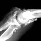
Synovialchondromatose
Kniegelenk. Zusätzlich ausgeprägte lateral betonte, deformierende Gonarthrose. Weiterhin auch Retropatellararthrose und Stieda-Pellegrini-Schatten.
Synovial chondromatosis (osteochondromatosis or synovial chondrometaplasia) also known as Reichel syndrome, is a disorder characterized by loose cartilaginous bodies which may, or may not be calcified or ossified.
It is classified under two main types:
Siehe auch:
- tumoröse Kalzinose
- Pigmentierte villonoduläre Synovialitis
- Lipoma arborescens
- siderotic synovitis
- freier Gelenkkörper
- sekundäre synoviale Chondromatose
- synoviale Raumforderungen
- Synovialchondromatose Hüftgelenk
- giant solitary synovial chondromatosis
- synoviales Hämangiohamartom
- synoviale Osteochondromatose Schultergelenk
- Synovialchondromatose Kniegelenk
und weiter:
- Weichteilverkalkungen
- Rice bodies (musculoskeletal)
- Renale Osteodystrophie
- Bursa subacromialis
- inanimate object inspired signs
- Riesenzelltumor der Sehnenscheiden
- fruit inspired signs
- extraskeletal musculoskeletal tumors by compartment
- Chondromatose
- differential diagnosis for calcified masses in the mandible
- Gelenktumoren
- anterior hip pain
- Coxa saltans
- extra skeletal musculoskeletal lesions by compartment
- Osteochondromatose
- rice bodies rheumatoide Arthritis
- Synoviale Chondromatose Kiefergelenk
- sarcoma fusigiganocellulare
- synoviale Chondromatose Ellenbogen
- polymorphocellular tumour of the synovial membrane
- hämophile Arthropathie
- Lipoma arborescens Kniegelenk
- chronic hemorrhagic villous synovitis
- synovial fibroendothelioma
- myeloplaxoma
- giant cell fibrohemangioma
- fibrohemosideric sarcoma
- synoviale Osteochondromatose bei Kindern
- synoviales Chondrom
- Apfelbutzenzeichen Femur
- synovial osteochondromatosis: coracoid bursa
- Hämangiom der Synovialmembran
- synovial chondromatosis of the shoulder
- synoviale Chondromatose Fuß
- primary synovial chondromatosis of the wrist joint
- synovial chondromatosis of elbow
- distribution of synovial osteochondromatosis
- doppellagige Patella
- synovial osteochondromatosis of the ankle
- Merkspruch Weichteilverkalkungen
- synoviale Osteochondromatose Ellenbogen


 Assoziationen und Differentialdiagnosen zu synoviale Osteochondromatose:


synoviale
Osteochondromatose Schultergelenk