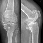
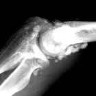
primary synovial chondromatosis


Primary synovial chondromatosis (also known as Reichel syndrome or Reichel-Jones-Henderson syndrome), is a benign monoarticular disorder of unknown origin that is characterized by synovial metaplasia and proliferation resulting in multiple intra-articular cartilaginous loose bodies of relatively similar size, not all of which are ossified. Hence, the term synovial chondromatosis is preferred over primary synovial osteochondromatosis. It is distinct from secondary synovial chondromatosis that is the result of a degenerative change in the joint.
Epidemiology
The age range of affected patients is wide, but most present in the 4 or 5 decades of life. Men are affected more frequently (M:F ratio of 2:1 to 4:1) .
Clinical presentation
Patients usually present with pain, swelling, and limitation of motion, which often progresses slowly for several years. Joint effusions are common as there is a restricted range of motion.
Pathology
Primary synovial chondromatosis is a self-limiting benign neoplastic process characterized by proliferative chondroid nodules of the synovium. Three phases of articular disease have been identified:
Location
Usually, the condition is monoarticular affecting any joint but the large joints are preferentially affected:
- knee (up to 70%)
- hip (20%)
- elbow
- shoulder
Occasionally, bursa or tendon sheaths may be involved .
Macroscopic appearance
Macroscopic appearance is that of a multilobulated synovium with multiple white/bluish nodules that are composed of hyaline cartilage attached to the synovium. These nodules may detach to form loose bodies. Most nodules are small (less than 2-3 cm) and usually uniform in size. Cases of massive nodules have been reported, with multiple nodules coalescing into giant nodules measuring up to 20 cm in size .
Histology
Microscopically the metaplastic synovium demonstrates cartilaginous nodules beneath the surface lining of the synovial membrane. They are characterized by proliferation and metaplastic transformation of the synovium, with formation of multiple cartilaginous or osteocartilaginous nodules within the joints, bursae, or tendon sheaths. These nodules are highly cellular, and the moderate pleomorphism may be identified. Cartilaginous bodies may contain cartilage alone, cartilage and bone, or mature bone with fatty marrow.
Radiographic features
Imaging findings depend on the stage of disease and the extent of calcification or ossification of the cartilaginous nodules. In its most distinctive appearance, multiple small, well-defined, juxta-articular nodules of uniform size are observed.
Plain radiograph and CT
The radiographic features depend on the degree of ossification that has occurred. When calcification is absent (25-30% of cases) plain radiographs may be normal or reveal a non-specific findings, e.g. soft-tissue mass surrounding the joint, widening of the joint space, erosions of adjacent bones, or early osteoarthritic changes.
When extensive ossification is present, then many calcific joint bodies are present, either fully ossified, or demonstrating the ring and arc calcification characteristic of chondroid calcifications. They are most often multiple and of uniform size .
CT may be able to confirm that the loose bodies are intra-articular, and arthrography can be used as an adjunct.
MRI
MRI appearance is variable and depends on the relative preponderance of synovial proliferation, loose bodies formation, and extent of calcification or ossification.
The most frequent pattern is one of predominantly unmineralised nodules that demonstrate typical chondroid signal characteristics:
- T1: intermediate to low signal
- T2: high signal
Focal areas of signal void within these nodules represent areas of mineralization .
- gradient echo (GE): will show blooming artefact
In some cases no mineralization is present, and in other instances (representing 'burnt out' disease) all the nodules are fully ossified with central fat intensity in keeping with marrow.
Treatment and prognosis
Treatment of synovial chondromatosis usually consists of removal of the intra-articular bodies with or without synovectomy, but local recurrence is not uncommon, occurring in ~12.5% (range 3-23%) of cases .
Malignant degeneration into chondrosarcoma has been reported but is rare . Additionally, the cellular atypia demonstrated as synovial osteochondromatosis may be misinterpreted in some instances as chondrosarcoma, and thus a true rate of malignant degeneration is uncertain .
History and etymology
Friedrich Paul Reichel (1858-1934) was a German surgeon who published the first description of this entity (in the German language) .
Hugh Toland Jones (1892-fl.1964) and Melvin Starkey Henderson (1883-1964) were both American orthopedic surgeons who wrote early publications on this condition .
Differential diagnosis
The differential diagnosis of primary synovial chondromatosis includes:
- secondary osteochondromatosis
- older age group
- extensive degenerative change
- fragments are fewer and often larger
- pigmented villonodular synovitis (PVNS)
- more confluent masses
- diffuse characteristic low intensity on MRI
- synovial hemangioma
- lipoma arborescens
- characteristic fat signal/density
- synovial chondrosarcoma
- soft tissue mass
- extension beyond the joint
- presence of metastases
- siderotic synovitis
- tumoral calcinosis
- peri-articular melorheostosis
See also
Siehe auch:
- tumoröse Kalzinose
- Pigmentierte villonoduläre Synovialitis
- Lipoma arborescens
- siderotic synovitis
- freier Gelenkkörper
- sekundäre synoviale Chondromatose
- synoviale Raumforderungen
- Synovialchondromatose Hüftgelenk
- giant solitary synovial chondromatosis
- synoviale Osteochondromatose Schultergelenk
- synoviales Hämangiohamartom
- Synovialchondromatose Kniegelenk
und weiter:
- Weichteilverkalkungen
- Rice bodies (musculoskeletal)
- Renale Osteodystrophie
- Bursa subacromialis
- inanimate object inspired signs
- Riesenzelltumor der Sehnenscheiden
- fruit inspired signs
- extraskeletal musculoskeletal tumors by compartment
- differential diagnosis for calcified masses in the mandible
- Chondromatose
- Gelenktumoren
- anterior hip pain
- Coxa saltans
- extra skeletal musculoskeletal lesions by compartment
- Synoviale Chondromatose Kiefergelenk
- rice bodies rheumatoide Arthritis
- Osteochondromatose
- sarcoma fusigiganocellulare
- polymorphocellular tumour of the synovial membrane
- synoviale Chondromatose Ellenbogen
- hämophile Arthropathie
- Lipoma arborescens Kniegelenk
- chronic hemorrhagic villous synovitis
- synovial fibroendothelioma
- giant cell fibrohemangioma
- myeloplaxoma
- fibrohemosideric sarcoma
- synoviale Osteochondromatose bei Kindern
- synoviales Chondrom
- synovial osteochondromatosis: coracoid bursa
- Apfelbutzenzeichen Femur
- Hämangiom der Synovialmembran
- synovial chondromatosis of the shoulder
- synoviale Chondromatose Fuß
- primary synovial chondromatosis of the wrist joint
- synovial chondromatosis of elbow
- distribution of synovial osteochondromatosis
- doppellagige Patella
- synovial osteochondromatosis of the ankle
- Merkspruch Weichteilverkalkungen
- synoviale Osteochondromatose Ellenbogen

 Assoziationen und Differentialdiagnosen zu synoviale Osteochondromatose:


