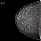
asymmetrische mammographische Dichte

Asymmetries in mammography represent a spectrum of morphological descriptors for a unilateral fibroglandular-density finding seen on one or more mammographic projections that do not meet criteria for a mass. The term refers to a density finding and should not be confused with asymmetry in breast size.
The criteria for an asymmetry include that it is seen only on one projection, the borders are not convex, or the center is not denser than the periphery (e.g. it is interspersed with fat).
Under the BI-RADS lexicon , there are four types of asymmetries:
- asymmetry
- focal asymmetry
- developing asymmetry
- global asymmetry
Pathology
The most common cause for an asymmetry on screening mammography is superimposition of normal breast tissue (summation artifact) . Asymmetries that are subsequently confirmed to be a real lesion may represent a focal asymmetry or mass, for which it is important to further evaluate to exclude breast cancer . Developing asymmetries are sufficiently suspicious to justify recall and biopsy, with 15% representing malignancy .
Global asymmetry is most commonly a normal variant and is discussed separately.
Radiographic features
The BI-RADS lexicon defines four types of asymmetries :
- asymmetry: visible on only one projection
- focal asymmetry: visible on two projections, involves less than one quadrant, lacks convex-outwards borders or is interspersed with fat
- developing asymmetry: focal asymmetry that is new, larger, or more conspicuous than on prior examinations
- global asymmetry: visible on two projections, involves more than one quadrant
Mammography
An asymmetry or focal asymmetry that is unchanged over at least 2 years does not deserve attention. Otherwise, findings of an asymmetry, focal asymmetry, or developing asymmetry found on screening merit recall for further evaluation.
Upon recall from screening mammography, repeating the original view(s) with the finding is often helpful and additional views should be considered:
- spot compression views: useful to distinguish superimposition of tissues from real findings (summation shadows disappear upon compression)
- rolled CC view: useful to localize a finding only seen on CC view
- 90 degree lateral view/LM view: useful to localize a finding better seen on MLO projection
- spot magnification views: rarely helpful for asymmetries alone but useful for evaluation of associated microcalcifications
In the diagnostic setting, localized findings can be further evaluated by ultrasound.
Radiology report
Asymmetries that turn out to be summation artifact are benign (BI-RADS 2). The BI-RADS Atlas offers guidance regarding the other categories of asymmetries :
A solitary focal asymmetry (without architectural distortion, calcifications, or underlying mass identified on diagnostic mammography and ultrasound) is assessed as BI-RADS 3 (likely benign).
A developing asymmetry, unless shown to be characteristically benign such as a cyst on ultrasound, is assessed BI-RADS 4 (suspicious). An exception would be if there is a clear benign explanation, such as recent surgery, trauma, or infection at that site.
Global asymmetry, in the absence of palpable correlate, is assessed BI-RADS 2 (benign).
Differential diagnosis
Asymmetries may represent any of a long list of pathologies:
- normal variation
- asymmetrical breast tissue
- ectopic breast tissue: accessory breast tissue
- post-traumatic
- asymmetry of residual parenchyma post breast reduction surgery
- sclerosing lobular hyperplasia
- pseudoangiomatous stromal hyperplasia (PASH)
- diabetic mastopathy
- breast cancer
See also
- other imaging features of breast malignancy
Siehe auch:
- invasives lobuläres Karzinom
- Liponekrosen Mamma
- Mamma accessoria
- Pseudoangiomatöse-Stroma-Hyperplasie (PASH)
- fibroadenomatoid mastopathy
- diabetische Mastopathie
- Lymphom der Mamma
- tubular carcinoma of breast
- global asymmetry in breast tissue
- postoperative Narben Mamma
- Neoplasien der Mamma
- step oblique mammography
- radiäre Narbe der Mamma
- Hämatom in der Mamma
- Anisomastie
- mammographic architectural distortion

 Assoziationen und Differentialdiagnosen zu asymmetrische mammographische Dichte:


