Calvarial doughnut lesion

Calvarial doughnut lesions are radiolucent ring-like skull defects, with surrounding sclerotic haloes, which may have central bone density, and may occur in any part of the skull.
Epidemiology
Most of these lesions occur in middle and old age, but also may be seen in juvenile skulls .
Clinical presentation
Calvarial doughnut lesions are usually an isolated incidental asymptomatic finding with no clinical significance .
Occasionally this entity forms part of a rare autosomal dominantly inherited syndrome, called familial calvarial doughnut lesions syndrome or calvarial doughnut lesions – bone fragility syndrome . This entity characterizes by multiple calvarial doughnut lesions, with lumps on the head, many pathologic fractures, dental caries, undeveloped teeth, and elevated serum alkaline phosphatase (ALP) levels .
Pathology
Microscopic features
These lesions may show central mesenchymal and fibrous tissue with clusters of foamy cells, usually with a nidus of irregular bone trabeculae and surrounding sclerotic cortical bone . There is no osteoblastic or osteoclastic proliferation; and no hemosiderin deposition, eosinophils, or inflammatory cells .
Etiology
The etiology of this entity remains unknown .
Radiographic features
Plain radiograph
The doughnut lesions occur in any portion of the skull and present a ring-like aspect, which may be divided into two groups by their radiographic configuration :
- group I: presents as a small, well-defined area of radiolucency, surrounded by a dense sclerotic bone, similar to doughnuts
- group II are usually larger than the first group, and the radiolucency and the sclerotic margins less regular
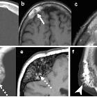
Lesions of both groups may contain areas of the density of varying sizes within the central areas of radiolucency . Some lesions may expand the outer table without bone erosion, usually corresponding to the area of the patient lump at skull .
CT
CT shows doughnut lesions within the diploe consisting of a round radiolucency lesion surrounded by a well-defined sclerotic ring, which may have irregular central sclerosis . Some lesions may be associated with bulging of the outer table , which usually corresponds to the area of the patient's skull lump.
MRI
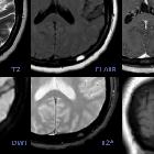
MR reveals expansile diploic homogeneous well-perfused soft-tissue lesions low signal intensity in T2 WI due to the high content of fibrous tissue . There is a hypointense ringlike structure surrounding these lesions, which represents the sclerotic ring . The CT is best to see the calcified portion of the lesion .
Treatment and prognosis
There is currently no evidence for intervention or surgical management of the disorder .
History and etymology
Keats and Holt described calvarial doughnut lesions for the first time in 1969 .
Differential diagnosis
It is essential to recognize these lesions as a benign process and to distinguish them from other lytic skull lesions :
- vascular markings
- intradiploic epidermoid cyst
- hemangioma
- eosinophilic granuloma
- lytic skeletal metastasis
Siehe auch:

 Assoziationen und Differentialdiagnosen zu Doughnut Zeichen Schädelkalotte:


