intradiploic epidermoid cyst

Intradiploic
epidermoid cyst with osteolytic lesions of the skull.. Computed tomography scan with bone density window reveal an occipital osteolytic lesion (arrows).

Intradiploic
epidermoid cyst • Skull vault epidermoid - Ganzer Fall bei Radiopaedia

Intradiploic
epidermoid cyst • Intradiploic epidermoid cyst - Ganzer Fall bei Radiopaedia

Intradiploic
epidermoid cyst • Large intradiploic epidermoid cyst - Ganzer Fall bei Radiopaedia

Intradiploic
epidermoid cyst • Intradiploic epidermoid cyst - Ganzer Fall bei Radiopaedia

Intradiploic
epidermoid cyst • Orbital intradiploic epidermoid cyst causing proptosis - Ganzer Fall bei Radiopaedia

Intradiploic
epidermoid cyst • Angular epidermoid cyst - Ganzer Fall bei Radiopaedia

Intradiploic
epidermoid cyst • Intradiploic epidermoid cyst - Ganzer Fall bei Radiopaedia

Intradiploic
epidermoid cyst • Intradiploic epidermoid - skull - Ganzer Fall bei Radiopaedia

Intradiploic
epidermoid cyst with osteolytic lesions of the skull.. Axial T2-weighted images demonstrate an inhomogeneous hyperintense lesion in the right occipital intradiploic space (arrows).

Intradiploic
epidermoid cyst • Intradiploic epidermoid - skull - Ganzer Fall bei Radiopaedia
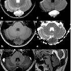
Spectrum of
lytic lesions of the skull: a pictorial essay. IEC. On MR images of the same patient shown in Fig. 3, the lesion appears slightly inhomogeneous, mostly hyperintense on T2w (a) and hypointense on T1w (b) images, without contrast enhancement (c), showing diffusion restriction on b 1000 diffusion-weighted imaging (DWI) and the apparent diffusion coefficient (ADC) map (d, e). It causes compression on the confluence of sinuses (sagittal contrast-enhanced T1w image, f). As a consequence, the patient presented with intracranial hypertension symptoms

Spectrum of
lytic lesions of the skull: a pictorial essay. Intradiploic epidermoid cyst (IEC). Axial CT images with soft tissues (a) and bone (b) windows show an occipital hypodense osteolytic lesion with smooth margins and greater involvement of the inner table, extensively demineralised

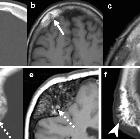
Radiological
review of skull lesions. Epidermoid cyst. Axial head CT portrays an expansile lesion in the right frontal bone (thick arrow) with thinning of the inner and outer tables (dashed arrows). Axial T1-weighted imaging (b), axial T2-weighted imaging (c), axial DWI (d) and axial post contrast T1-weighted imaging (e) show that the lesion is hypointense on T1, hyperintense on T2 with restricted diffusion and absent internal enhancement (arrowheads). Note that there may be some enhancement along the peripheral margins

Intradiploic
epidermoid cyst with osteolytic lesions of the skull.. The ADC maps reveal restricted diffusion (arrows).

Intradiploic
epidermoid cyst with osteolytic lesions of the skull.. Axial post gadolinium T1-weighted MR images show minimal and peripheral contrast enhancement (arrows).

Intradiploic
epidermoid cyst with osteolytic lesions of the skull.. Axial FLAIR images demonstrate an inhomogeneous hyperintense lesion in the right occipital intradiploic space (arrows).

Intradiploic
epidermoid cyst • Intradiploic epidermoid cyst - Ganzer Fall bei Radiopaedia
Intradiploic epidermoid cysts represent epidermoid cysts that occur in the diploë of the skull.
Clinical presentation
Painless slowly progressive scalp swelling.
Pathology
- epidermoid cysts may be congenital (most common, arising from ectodermal inclusion during neural tube closure and subsequently remain within the cranial bones) or acquired (e.g. post-surgical or post-traumatic implantation)
- intradiploic epidermoids are less frequent than the intradural variety
Radiographic features
Intradiploic epidermoids occurs within the frontal, parietal, occipital and sphenoid bones, as well as the spine .
Plain radiograph
- rounded or lobulated area of bone destruction, well-delineated sclerotic scalloped margins
CT
- non-enhancing hypodense lesion with sharply demarcated bony defects and zones of calcifications
- it may alter the outer and/or inner tables of the skull (the inner table more than the outer)
MRI
- T1: slightly hyperintense to the CSF
- T2: isointense/hyperintense to the CSF
- FLAIR: hyperintense to the CSF space
- DWI: restricted diffusion with characteristic hyperintensity
- T1C+: none
History and etymology
The first intradiploic epidermoid cyst was reported by J Müller in 1838 .
The radiological pattern of intradiploic epidermoids was first described by Cushing in 1922.
Differential diagnosis
Consider:
- eosinophilic granuloma
- calvarial hemangioma
- dermoid cyst
- fibrous dysplasia
Siehe auch:


 Assoziationen und Differentialdiagnosen zu Epidermoidzyste der Kalotte: